12
views
0
recommends
+1 Recommend
0 collections
    0
    shares
      • Record: found
      • Abstract: found
      • Article: found
      Is Open Access

      In vivo UVA irradiation of mouse is more efficient in promoting pulmonary melanoma metastasis than in vitro

      research-article

      Read this article at

      Bookmark
          There is no author summary for this article yet. Authors can add summaries to their articles on ScienceOpen to make them more accessible to a non-specialist audience.

          Abstract

          Background

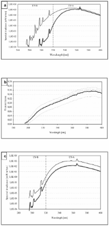
          We have previously shown in vitro that UVA increases the adhesiveness of mouse B16-F1 melanoma cells to endothelium.

          We have also shown in vivo that UVA exposure of C57BL/6 mice, i.v. injected with B16-F1 cells, increases formation of pulmonary colonies of melanoma. The aim of the present animal study was to confirm the previously observed in vivo UVA effect and to determine whether in vitro UVA-exposure of melanoma cells, prior the i.v. injection, will have an enhancing effect on the pulmonary colonization capacity of melanoma cells. As a second aim, UVA-derived immunosuppression was determined.

          Methods

          Mice were i.v. injected with B16-F1 cells into the tail vein and then immediately exposed to UVA. Alternatively, to study the effect of UVA-induced adhesiveness on the colonization capacity of B16-F1 melanoma, cells were in vitro exposed prior to i.v. injection. Fourteen days after injection, lungs were collected and the number of pulmonary nodules was determined under dissecting microscope. The UVA-derived immunosuppression was measured by standard contact hypersensitivity assay.

          Results and Discussion

          Obtained results have confirmed that mice, i.v. injected with B16-F1 cells and thereafter exposed to UVA, developed 4-times more of melanoma colonies in lungs as compared with the UVA non-exposed group (p < 0.01). The in vitro exposure of melanoma cells prior to their injection into mice, led only to induction of 1.5-times more of pulmonary tumor nodules, being however a statistically non-significant change. The obtained results postulate that the UVA-induced changes in the adhesive properties of melanoma cells do not alone account for the 4-fold increase in the pulmonary tumor formation. Instead, it suggests that some systemic effect in a mouse might be responsible for the increased metastasis formation. Indeed, UVA was found to induce moderate systemic immunosuppression, which effect might contribute to the UVA-induced melanoma metastasis in mice lungs.

          Related collections

          Most cited references27

          • Record: found
          • Abstract: not found
          • Article: not found

          Selection of successive tumour lines for metastasis.

          I J Fidler (1973)
            Bookmark
            • Record: found
            • Abstract: found
            • Article: not found

            Wavelength dependence of oxidative DNA damage induced by UV and visible light.

            DNA damage induced by UV radiation and visible light (290-500 nm) in AS52 Chinese hamster cells was analysed by an alkaline elution assay with specific repair endonucleases. Cells were exposed to extensively filtered monochrome or broad-band radiation. Between 290 and 315 nm, the ratio of base modifications sensitive to Fpg protein (i.e. 8-hydroxyguanine and formamidopyrimidines) and T4 endonuclease V (i.e. cyclobutane pyrimidine dimers) was constant (approximately 1:200), indicating that the direct excitation of DNA is responsible for both types of damage in this range of the spectrum. While the yield of pyrimidine dimers per unit dose continued to decrease exponentially beyond 315 nm, the yield of Fpg-sensitive modifications increased to a second maximum between 400 and 450 nm. The damage spectrum in this wavelength range consisted of only a few other modifications (strand breaks, abasic sites and pyrimidine modifications sensitive to endonuclease III) and is attributed to endogenous photosensitizers that give rise to oxidative DNA damage via singlet oxygen and/or type I reactions. The generation of Fpg-sensitive modifications by visible light was not linear with dose but followed a saturation curve. It is calculated that the exposure of the cells to low doses of solar radiation results in the formation of cyclobutane pyrimidine dimers and Fpg-sensitive modifications in a ratio of 10:1.
              Bookmark
              • Record: found
              • Abstract: found
              • Article: not found

              The basal layer in human squamous tumors harbors more UVA than UVB fingerprint mutations: a role for UVA in human skin carcinogenesis.

              We hypothesized that a substantial portion of the mutagenic alterations produced in the basal layer of human skin by sunlight are induced by wavelengths in the UVA range. Using laser capture microdissection we examined separately basal and suprabasal keratinocytes from human skin squamous cell carcinomas and premalignant solar keratosis for both UVA- and UVB-induced adduct formation and signature mutations. We found that UVA fingerprint mutations were detectable in human skin squamous cell carcinomas and solar keratosis, mostly in the basal germinative layer, which contrasted with a predominantly suprabasal localization of UVB fingerprint mutations in these lesions. The epidermal layer bias was confirmed by immunohistochemical analyses with a superficial localization of cyclobutane thymine dimers contrasting with the localization of 8-hydroxy-2'-deoxyguanine adducts to the basal epithelial layers. If unrepaired, these adducts may lead to fixed genomic mutations. The basal location of UVA-rather than UVB-induced DNA damage suggests that longer-wavelength UVR is an important carcinogen in the stem cell compartment of the skin. Given the traditional emphasis on UVB, these results may have profound implications for future public health initiatives for skin cancer prevention.
                Bookmark

                Author and article information

                Journal
                Cancer Cell Int
                Cancer Cell International
                BioMed Central
                1475-2867
                2011
                6 June 2011
                : 11
                : 16
                Affiliations
                [1 ]Non-Ionizing Radiation Laboratory, STUK-Radiation and Nuclear Safety Authority, Laippatie 4, FIN-00880, Helsinki, Finland
                [2 ]Health Risks and Radon Safety Laboratory; STUK-Radiation and Nuclear Safety Authority, Laippatie 4, FIN-00880, Helsinki, Finland
                [3 ]Radiation Biology Laboratory; STUK-Radiation and Nuclear Safety Authority, Laippatie 4, FIN-00880, Helsinki, Finland
                Article
                1475-2867-11-16
                10.1186/1475-2867-11-16
                3123265
                21645404
                0be7661a-8060-4208-9452-78ccd85ecf7a
                Copyright ©2011 Pastila et al; licensee BioMed Central Ltd.

                This is an Open Access article distributed under the terms of the Creative Commons Attribution License ( http://creativecommons.org/licenses/by/2.0), which permits unrestricted use, distribution, and reproduction in any medium, provided the original work is properly cited.

                History
                : 10 January 2011
                : 6 June 2011
                Categories
                Primary Research

                Oncology & Radiotherapy
                immunosuppression,mouse melanoma,uva radiation,b16,metastasis
                Oncology & Radiotherapy
                immunosuppression, mouse melanoma, uva radiation, b16, metastasis

                Comments

                Comment on this article