38
views
0
recommends
+1 Recommend
0 collections
    0
    shares
      • Record: found
      • Abstract: found
      • Article: found
      Is Open Access

      Treatment with Riluzole Restores Normal Control of Soleus and Extensor Digitorum Longus Muscles during Locomotion in Adult Rats after Sciatic Nerve Crush at Birth

      research-article
      1 , * , 1 , 2 , 2
      PLoS ONE
      Public Library of Science

      Read this article at

      Bookmark
          There is no author summary for this article yet. Authors can add summaries to their articles on ScienceOpen to make them more accessible to a non-specialist audience.

          Abstract

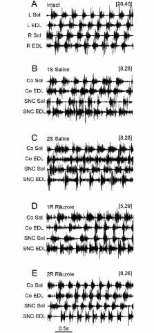
          The effects of sciatic nerve crush (SNC) and treatment with Riluzole on muscle activity during unrestrained locomotion were identified in an animal model by analysis of the EMG activity recorded from soleus (Sol) and extensor digitorum longus (EDL) muscles of both hindlimbs; in intact rats (IN) and in groups of rats treated for 14 days with saline (S) or Riluzole (R) after right limb nerve crush at the 1 st (1S and 1R) or 2 nd (2S and 2R) day after birth. Changes in the locomotor pattern of EMG activity were correlated with the numbers of survived motor units (MUs) identified in investigated muscles. S rats with 2–8 and 10–28 MUs that survived in Sol and EDL muscles respectively showed increases in the duration and duty factor of muscle EMG activity and a loss of correlation between the duty factors of muscle activity, and abnormal flexor-extensor co-activation 3 months after SNC. R rats with 5, 6 (Sol) and 15–29 MUs (EDL) developed almost normal EMG activity of both Sol and control EDL muscles, whereas EDL muscles with SNC showed a lack of recovery. R rats with 8 (Sol) and 23–33 (EDL) MUs developed almost normal EMG activities of all four muscles. A subgroup of S rats with a lack of recovery and R rats with almost complete recovery that had similar number of MUs (8 and 24–28 vs 8 and 23–26), showed that the number of MUs was not the only determinant of treatment effectiveness. The results demonstrated that rats with SNC failed to develop normal muscle activity due to malfunction of neuronal circuits attenuating EDL muscle activity during the stance phase, whereas treatment with Riluzole enabled almost normal EMG activity of Sol and EDL muscles during locomotor movement.

          Related collections

          Most cited references31

          • Record: found
          • Abstract: found
          • Article: not found

          Dynamic sensorimotor interactions in locomotion.

          Locomotion results from intricate dynamic interactions between a central program and feedback mechanisms. The central program relies fundamentally on a genetically determined spinal circuitry (central pattern generator) capable of generating the basic locomotor pattern and on various descending pathways that can trigger, stop, and steer locomotion. The feedback originates from muscles and skin afferents as well as from special senses (vision, audition, vestibular) and dynamically adapts the locomotor pattern to the requirements of the environment. The dynamic interactions are ensured by modulating transmission in locomotor pathways in a state- and phase-dependent manner. For instance, proprioceptive inputs from extensors can, during stance, adjust the timing and amplitude of muscle activities of the limbs to the speed of locomotion but be silenced during the opposite phase of the cycle. Similarly, skin afferents participate predominantly in the correction of limb and foot placement during stance on uneven terrain, but skin stimuli can evoke different types of responses depending on when they occur within the step cycle. Similarly, stimulation of descending pathways may affect the locomotor pattern in only certain phases of the step cycle. Section ii reviews dynamic sensorimotor interactions mainly through spinal pathways. Section iii describes how similar sensory inputs from the spinal or supraspinal levels can modify locomotion through descending pathways. The sensorimotor interactions occur obviously at several levels of the nervous system. Section iv summarizes presynaptic, interneuronal, and motoneuronal mechanisms that are common at these various levels. Together these mechanisms contribute to the continuous dynamic adjustment of sensorimotor interactions, ensuring that the central program and feedback mechanisms are congruous during locomotion.
            Bookmark
            • Record: found
            • Abstract: found
            • Article: not found

            Organization of mammalian locomotor rhythm and pattern generation.

            Central pattern generators (CPGs) located in the spinal cord produce the coordinated activation of flexor and extensor motoneurons during locomotion. Previously proposed architectures for the spinal locomotor CPG have included the classical half-center oscillator and the unit burst generator (UBG) comprised of multiple coupled oscillators. We have recently proposed another organization in which a two-level CPG has a common rhythm generator (RG) that controls the operation of the pattern formation (PF) circuitry responsible for motoneuron activation. These architectures are discussed in relation to recent data obtained during fictive locomotion in the decerebrate cat. The data show that the CPG can maintain the period and phase of locomotor oscillations both during spontaneous deletions of motoneuron activity and during sensory stimulation affecting motoneuron activity throughout the limb. The proposed two-level CPG organization has been investigated with a computational model which incorporates interactions between the CPG, spinal circuits and afferent inputs. The model includes interacting populations of spinal interneurons and motoneurons modeled in the Hodgkin-Huxley style. Our simulations demonstrate that a relatively simple CPG with separate RG and PF networks can realistically reproduce many experimental phenomena including spontaneous deletions of motoneuron activity and a variety of effects of afferent stimulation. The model suggests plausible explanations for a number of features of real CPG operation that would be difficult to explain in the framework of the classical single-level CPG organization. Some modeling predictions and directions for further studies of locomotor CPG organization are discussed.
              Bookmark
              • Record: found
              • Abstract: found
              • Article: not found

              Permanent central synaptic disconnection of proprioceptors after nerve injury and regeneration. I. Loss of VGLUT1/IA synapses on motoneurons.

              Motor and sensory proprioceptive axons reinnervate muscles after peripheral nerve transections followed by microsurgical reattachment; nevertheless, motor coordination remains abnormal and stretch reflexes absent. We analyzed the possibility that permanent losses of central IA afferent synapses, as a consequence of peripheral nerve injury, are responsible for this deficit. VGLUT1 was used as a marker of proprioceptive synapses on rat motoneurons. After nerve injuries synapses are stripped from motoneurons, but while other excitatory and inhibitory inputs eventually recover, VGLUT1 synapses are permanently lost on the cell body (75-95% synaptic losses) and on the proximal 100 μm of dendrite (50% loss). Lost VGLUT1 synapses did not recover, even many months after muscle reinnervation. Interestingly, VGLUT1 density in more distal dendrites did not change. To investigate whether losses are due to VGLUT1 downregulation in injured IA afferents or to complete synaptic disassembly and regression of IA ventral projections, we studied the central trajectories and synaptic varicosities of axon collaterals from control and regenerated afferents with IA-like responses to stretch that were intracellularly filled with neurobiotin. VGLUT1 was present in all synaptic varicosities, identified with the synaptic marker SV2, of control and regenerated afferents. However, regenerated afferents lacked axon collaterals and synapses in lamina IX. In conjunction with the companion electrophysiological study [Bullinger KL, Nardelli P, Pinter MJ, Alvarez FJ, Cope TC. J Neurophysiol (August 10, 2011). doi:10.1152/jn.01097.2010], we conclude that peripheral nerve injuries cause a permanent retraction of IA afferent synaptic varicosities from lamina IX and disconnection with motoneurons that is not recovered after peripheral regeneration and reinnervation of muscle by sensory and motor axons.
                Bookmark

                Author and article information

                Contributors
                Role: Editor
                Journal
                PLoS One
                PLoS ONE
                plos
                plosone
                PLoS ONE
                Public Library of Science (San Francisco, CA USA )
                1932-6203
                17 January 2017
                2017
                : 12
                : 1
                : e0170235
                Affiliations
                [1 ]Department of Engineering of Nervous and Muscular System, Nałęcz Institute of Biocybernetics and Biomedical Engineering, PAS, Warsaw, Poland
                [2 ]Department of Neurophysiology, Nencki Institute of Experimental Biology, PAS, Warsaw, Poland
                Szegedi Tudomanyegyetem, HUNGARY
                Author notes

                Competing Interests: The authors have declared that no competing interests exist.

                • Conceptualization: WZ AMC US.

                • Formal analysis: WZ.

                • Investigation: AMC US.

                • Methodology: WZ AMC US.

                • Supervision: WZ AMC.

                • Visualization: WZ.

                • Writing – original draft: WZ AMC US.

                Author information
                http://orcid.org/0000-0002-2435-0078
                Article
                PONE-D-16-40363
                10.1371/journal.pone.0170235
                5240973
                28095499
                0c20cb24-b397-4468-8c6a-1aac60453fbc
                © 2017 Zmysłowski et al

                This is an open access article distributed under the terms of the Creative Commons Attribution License, which permits unrestricted use, distribution, and reproduction in any medium, provided the original author and source are credited.

                History
                : 10 October 2016
                : 2 January 2017
                Page count
                Figures: 11, Tables: 1, Pages: 28
                Funding
                This work was supported by grants from the Polish Ministry of Science and High Education 1527/PO1/2006/31 and statutory donation from the Nencki Institute as well as the Nałęcz Institute.
                Categories
                Research Article
                Research and Analysis Methods
                Bioassays and Physiological Analysis
                Electrophysiological Techniques
                Muscle Electrophysiology
                Electromyography
                Biology and Life Sciences
                Biomechanics
                Biological Locomotion
                Biology and Life Sciences
                Physiology
                Biological Locomotion
                Medicine and Health Sciences
                Physiology
                Biological Locomotion
                Biology and Life Sciences
                Anatomy
                Nervous System
                Neuroanatomy
                Neural Pathways
                Medicine and Health Sciences
                Anatomy
                Nervous System
                Neuroanatomy
                Neural Pathways
                Biology and Life Sciences
                Neuroscience
                Neuroanatomy
                Neural Pathways
                Biology and Life Sciences
                Anatomy
                Nervous System
                Nerves
                Sciatic Nerves
                Medicine and Health Sciences
                Anatomy
                Nervous System
                Nerves
                Sciatic Nerves
                Research and Analysis Methods
                Bioassays and Physiological Analysis
                Muscle Analysis
                Biology and Life Sciences
                Anatomy
                Musculoskeletal System
                Medicine and Health Sciences
                Anatomy
                Musculoskeletal System
                Biology and Life Sciences
                Anatomy
                Musculoskeletal System
                Muscles
                Soleus Muscles
                Medicine and Health Sciences
                Anatomy
                Musculoskeletal System
                Muscles
                Soleus Muscles
                Medicine and Health Sciences
                Critical Care and Emergency Medicine
                Trauma Medicine
                Traumatic Injury
                Musculoskeletal Injury
                Custom metadata
                All relevant data are within the paper and its Supporting Information files.

                Uncategorized
                Uncategorized

                Comments

                Comment on this article