15
views
0
recommends
+1 Recommend
0 collections
    0
    shares
      • Record: found
      • Abstract: found
      • Article: found
      Is Open Access

      Time course of eosinophilic myocarditis visualized by CMR

      research-article

      Read this article at

      Bookmark
          There is no author summary for this article yet. Authors can add summaries to their articles on ScienceOpen to make them more accessible to a non-specialist audience.

          Abstract

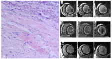
          We report the diagnostic potential of cardiovascular magnetic resonance (CMR) to visualize the time course of eosinophilic myocarditis upon successful treatment. A 50-year-old man was admitted with a progressive heart failure. Endomyocardial biopsies were taken from the left ventricle because of a white blood cell count of 17000/mm 3 with 41% eosinophils. Histological evaluation revealed endomyocardial eosinophilic infiltration and areas of myocyte necrosis. The patient was diagnosed with hypereosinophilic myocarditis due to idiopathic hypereosinophilic syndrome. CMR-studies at presentation and a follow-up study 3 weeks later showed diffuse subendocardial LGE in the whole left ventricle. Upon treatment with steroids, CMR-studies revealed marked reduction of subendocardial LGE after 3 months in parallel with further clinical improvement. This case therefore highlights the clinical importance of CMR to visualize the extent of endomyocardial involvement in the diagnosis and treatment of eosinophilic myocarditis.

          Related collections

          Most cited references5

          • Record: found
          • Abstract: not found
          • Article: not found

          Clinical and echocardiographic features of hypereosinophilic syndromes.

            Bookmark
            • Record: found
            • Abstract: found
            • Article: not found

            Ventricular remodeling in Loeffler endocarditis: implications for therapeutic decision making.

            Little is known about the morphological and functional evolution of ventricular abnormalities in Loeffler endocarditis. We describe 5 patients, including 3 with long-term echocardiographic follow-up, in whom apical obliteration due to fibro-thrombotic thickening of the endocardium showed favorable patterns of evolution. In one patient there was almost complete resolution of the obliterative process with consequently increased effective ventricular volume. In two patients formation of a flow-passage in the fibrocalcific apical 'floor' between the main medioventricular cavity and the apical chamber, leading to a 'double-chambered' left ventricle was observed. Medical therapy and appropriate anticoagulation, can induce favorable long-term ventricular remodeling in Loeffler endocarditis.
              Bookmark
              • Record: found
              • Abstract: not found
              • Article: not found

              Endocarditis parietalis fbroplastica mit Bluteosinophilie

                Bookmark

                Author and article information

                Journal
                J Cardiovasc Magn Reson
                Journal of Cardiovascular Magnetic Resonance
                BioMed Central
                1097-6647
                1532-429X
                2008
                8 May 2008
                : 10
                : 1
                : 21
                Affiliations
                [1 ]Klinik und Poliklinik für Innere Medizin II, Klinikum der Universität, Regensburg, Germany
                [2 ]Institut für Röntgendiagnostik, Klinikum der Universität, Regensburg, Germany
                Article
                1532-429X-10-21
                10.1186/1532-429X-10-21
                2413232
                18466607
                0e6da619-b790-4dd2-bb81-753d999766ed
                Copyright © 2008 Deb et al; licensee BioMed Central Ltd.

                This is an Open Access article distributed under the terms of the Creative Commons Attribution License ( http://creativecommons.org/licenses/by/2.0), which permits unrestricted use, distribution, and reproduction in any medium, provided the original work is properly cited.

                History
                : 26 March 2008
                : 8 May 2008
                Categories
                Case Report

                Cardiovascular Medicine
                Cardiovascular Medicine

                Comments

                Comment on this article