3
views
0
recommends
+1 Recommend
0 collections
    0
    shares
      • Record: found
      • Abstract: found
      • Article: found
      Is Open Access

      Fusion rates support wired allograft combined with instrumented craniocervical fixation in the paediatric population

      research-article

      Read this article at

      Bookmark
          There is no author summary for this article yet. Authors can add summaries to their articles on ScienceOpen to make them more accessible to a non-specialist audience.

          Abstract

          Background

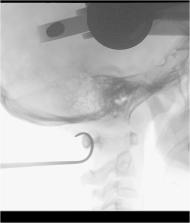
          Occipitocervical and atlantoaxial instability in the pediatric population is a rare and challenging condition to treat. Variable surgical techniques have been employed to achieve fusion. The study aimed to assess bony fusion with rigid craniocervical fixation using an allograft bone block to serve as scaffold for bony fusion.

          Methods

          This is a single center case series from a tertiary referral neurosurgical center. The series includes 12 consecutive pediatric patients with rigid craniocervical fusion between 2006 and 2014. The primary outcome was bony fusion as assessed by computed tomography and flexion-extension radiographs. The authors did not receive external funding for this study.

          Results

          Twelve patients (age 1–15 years) were operated with a median imaging follow-up time of 22 months (range 6–69 m). A modified Gallie fusion technique with a tightly wired allograft bone block was used in 10 of 13 procedures. One patient underwent re-fixation due to screw breakage. Eleven out of 13 procedures resulted in a stable construct with bony fusion. All 10 patients operated with the modified Gallie fusion technique with sublaminar wiring of allograft bone block had bony fusion. No post-operative complications of the posterior fixation procedure were noted.

          Conclusions

          The modified Gallie fusion technique with allograft bone block without post-operative immobilization achieved excellent fusion. We conclude there is no need to use autograft or BMPs in craniocervical fusion in the pediatric population, which avoids related donor-site morbidity.

          Level of evidence

          Level IV—case series; therapeutic.

          Related collections

          Most cited references19

          • Record: found
          • Abstract: found
          • Article: not found

          Healos and bone marrow aspirate used for lumbar spine fusion: a case controlled study comparing healos with autograft.

          A prospective case controlled study to compare the clinical and radiographic performance of Healos soaked in bone marrow aspirate (BMA) to iliac crest autograft when used in lumbar spinal fusion. To evaluate the null hypothesis: Healos used with BMA is not an effective alternative to iliac crest autograft in lumbar spine fusions. Healos (a Type 1 collagen/hydroxyapatite matrix) is osteoconductive and when soaked for at least 20 minutes in BMA becomes osteoinductive. It is nontoxic and straightforward to use, avoiding the morbidity of autograft harvest. Animal studies and early clinical series in humans have suggested that Healos and BMA are an effective substitute for autograft in certain circumstances. From July 2000, Healos and BMA were used as the graft material, instead of autograft harvested from the iliac crest, in all patients undergoing lumbar spinal fusion. Clinical outcome measures used were the Low Back Outcome Score (LBOS), a Patient Satisfaction Score, and the Prolo Economic Score (after Schnee). Standing anteroposterior and lateral radiographs were taken at 12- and 24-month follow-up visits. The first 50 cases in this consecutive series were age, sex, and operative intervention matched to historical controls who underwent surgery between 1997 and 2000 and in whom autograft from the iliac crest had been used as the graft material. Surgical outcome data in these patients had also been gathered prospectively. An independent radiologist, blinded to the graft material, using standard plain radiograph criteria for fusion, examined all the radiographs. An independent surgeon assessed clinical outcomes. For posterolateral lumbar fusions, there were equivalent radiologic fusion rates for the 2 groups with no significant difference in the subjective and objective clinical outcomes. The radiologic fusions rate was significantly lower when Healos had been used for lumbar interbody fusions. Clinical outcomes for both groups were similar. There were no lasting complications associated with Healos use compared with a 14%; persisting donor site complication rate in the autograft patients. The null hypothesis is only partially correct. Healos and BMA are not inferior to autologous iliac crest bone as a graft material in posterolateral lumbar spine fusions but are radiographically ineffective in lumbar interbody fusions.
            Bookmark
            • Record: found
            • Abstract: found
            • Article: not found

            Spine fusion using cell matrix composites enriched in bone marrow-derived cells.

            Bone marrow-derived cells including osteoblastic progenitors can be concentrated rapidly from bone marrow aspirates using the surface of selected implantable matrices for selective cell attachment. Concentration of cells in this way to produce an enriched cellular composite graft improves graft efficacy. The current study was designed to test the hypothesis that the biologic milieu of a bone marrow clot will significantly improve the efficacy of such a graft. An established posterior spinal fusion model and cancellous bone matrix was used to compare an enriched cellular composite bone graft alone, bone matrix plus bone marrow clot, and an enriched bone matrix composite graft plus bone marrow clot. Union score, quantitative computed tomography, and mechanical testing were used to define outcome. The union score for the enriched bone matrix plus bone marrow clot composite was superior to the enriched bone matrix alone and the bone matrix plus bone marrow clot. The enriched bone matrix plus bone marrow clot composite also was superior to the enriched bone matrix alone in fusion volume and in fusion area. These data confirm that the addition of a bone marrow clot to an enriched cell-matrix composite graft results in significant improvement in graft performance. Enriched composite grafts prepared using this strategy provide a rapid, simple, safe, and inexpensive method for intraoperative concentration and delivery of bone marrow-derived cells and connective tissue progenitors that may improve the outcome of bone grafting.
              Bookmark
              • Record: found
              • Abstract: found
              • Article: not found

              CT-based classification of long spinal allograft fusion.

              Anterior column reconstruction of the thoracolumbar spine by structural allograft has an increased potential for biological fusion when compared to synthetic reconstructive options. Estimation of cortical union and trabecular in-growth is, however, traditionally based on plain radiography, a technique lacking in sensitivity. A new assessment method of bony union using high-speed spiral CT imaging is proposed which reflects the gradually increasing biological stability of the construct. Grade I (complete fusion) implies cortical union of the allograft and central trabecular continuity. Grade II (partial fusion) implies cortical union of the structural allograft with partial trabecular incorporation. Grade III (unipolar pseudarthrosis) denotes superior or inferior cortical non-union of the central allograft with partial trabecular discontinuity centrally and Grade IV (bipolar pseudarthrosis) suggests both superior and inferior cortical non-union with a complete lack of central trabecular continuity. Twenty-five patients underwent anterior spinal reconstruction for a single level burst fracture between T4 and L5. At a minimum of two years follow up the subjects underwent high-speed spiral CT scanning through the reconstructed region of the thoracolumbar spine. The classification showed satisfactory interobserver (kappa score = 0.91) and intraobserver (kappa score = 0.95) reliability. The use of high-speed CT imaging in the assessment of structural allograft union may allow a more accurate assessment of union. The classification system presented allows a reproducible categorization of allograft incorporation with implications for treatment.
                Bookmark

                Author and article information

                Contributors
                w.pondaag@lumc.nl
                Journal
                Acta Neurochir (Wien)
                Acta Neurochir (Wien)
                Acta Neurochirurgica
                Springer Vienna (Vienna )
                0001-6268
                0942-0940
                24 March 2020
                24 March 2020
                2020
                : 162
                : 5
                : 985-991
                Affiliations
                [1 ]GRID grid.10419.3d, ISNI 0000000089452978, Department of Neurosurgery, , Leiden University Medical Center, ; PO Box 9600, 2300RC Leiden, The Netherlands
                [2 ]GRID grid.414842.f, ISNI 0000 0004 0395 6796, Department of Neurosurgery, , Haaglanden Medical Center, ; The Hague, The Netherlands
                Article
                4287
                10.1007/s00701-020-04287-9
                7156351
                32211968
                0eb326e6-0bcb-40e9-996a-2a57adda6061
                © The Author(s) 2020

                Open Access This article is licensed under a Creative Commons Attribution 4.0 International License, which permits use, sharing, adaptation, distribution and reproduction in any medium or format, as long as you give appropriate credit to the original author(s) and the source, provide a link to the Creative Commons licence, and indicate if changes were made. The images or other third party material in this article are included in the article's Creative Commons licence, unless indicated otherwise in a credit line to the material. If material is not included in the article's Creative Commons licence and your intended use is not permitted by statutory regulation or exceeds the permitted use, you will need to obtain permission directly from the copyright holder. To view a copy of this licence, visit http://creativecommons.org/licenses/by/4.0/.

                History
                : 15 November 2019
                : 10 March 2020
                Funding
                Funded by: Leiden University Medical Center (LUMC)
                Categories
                Original Article - Pediatric Spine
                Custom metadata
                © Springer-Verlag GmbH Austria, part of Springer Nature 2020

                Surgery
                allograft ,bone fusion ,children ,craniocervical instability ,surgical technique
                Surgery
                allograft , bone fusion , children , craniocervical instability , surgical technique

                Comments

                Comment on this article