2
views
0
recommends
+1 Recommend
0 collections
    0
    shares
      • Record: found
      • Abstract: found
      • Article: found
      Is Open Access

      Subclavian Artery Calcification: A Narrative Review

      review-article

      Read this article at

      Bookmark
          There is no author summary for this article yet. Authors can add summaries to their articles on ScienceOpen to make them more accessible to a non-specialist audience.

          Abstract

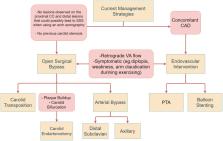
          Subclavian artery calcification (SAC) affects 2% of the population and presents a serious risk of developing into subclavian steal syndrome (SSS). Risk factors for plaque formation of the subclavian artery include diabetes, hypertension, and smoking. While SAC generally presents as asymptomatic, symptoms in severe cases may include numbness, pain at rest, and ischemic gangrene. Patients with severe SSS are at high risk of developing neurological symptoms as a result of vertebrobasilar insufficiency affecting posterior cerebral perfusion. On physical examination, SSS is preliminarily diagnosed from bilateral inter-arm systolic blood pressure discrepancy (>10 mmHg), which can be further confirmed with vascular imaging. Duplex ultrasound (DUS) is a cost-effective and non-invasive baseline technique for visualizing luminal stenosis and quantifying peak systolic velocity (PSV). Computed tomography angiography (CTA) provides high-quality, fast, three-dimensional (3D) imaging at the cost of introducing nephrotoxic contrast agents. Magnetic resonance angiography (MRA) is the safest 3D imaging modality, without the use of X-rays and contrast agents, that is useful in assessing plaque characteristics and degree of stenosis. DUS-assisted digital subtraction angiography (DSA) remains the gold standard for grading the degree of stenosis in the subclavian artery and determining the distance between the puncture site and lesion, which can be carried out in a combined procedure with endovascular management strategies. The fundamental treatment options are surgical and endovascular intervention. Endovascular treatment options include percutaneous transluminal angiography (PTA) for recanalization of the stenosed vessel and permanent balloon stenting to prevent collapse after PTA. Overall, the benefits of endovascular management encompass faster recovery, lower stenosis recurrence rate, and lower incidence of complications, making it the treatment of choice in low-risk patients. Surgical interventions, although more complex, are considered gold-standard treatment options.

          Related collections

          Most cited references50

          • Record: found
          • Abstract: found
          • Article: not found

          Vascular calcification: an update on mechanisms and challenges in treatment.

          Vascular calcification is highly associated with cardiovascular disease mortality, particularly in high-risk patients with diabetes and chronic kidney diseases (CKD). In blood vessels, intimal calcification is associated with atherosclerosis, whereas medial calcification is a nonocclusive process which leads to increased vascular stiffness and reduced vascular compliance. In the valves, calcification of the leaflets can change the mechanical properties of the tissue and result in stenosis. For many decades, vascular calcification has been noted as a consequence of aging. Studies now confirm that vascular calcification is an actively regulated process and shares many features with bone development and metabolism. This review provides an update on the mechanisms of vascular calcification including the emerging roles of the RANK/RANKL/OPG triad, osteoclasts, and microRNAs. Potential treatments adapted from osteoporosis and CKD treatments that are under investigation for preventing and/or regressing vascular calcification are also reviewed.
            Bookmark
            • Record: found
            • Abstract: found
            • Article: not found

            Coronary risk factors measured in childhood and young adult life are associated with coronary artery calcification in young adults: the Muscatine Study.

            This study was designed to estimate the prevalence of coronary artery calcification in young adult men and women and to examine the association between the presence of coronary artery calcification and coronary risk factors measured in childhood and young adult life. Electron beam computed tomography is a sensitive, noninvasive method for detecting coronary artery calcification, a marker of the atherosclerotic process. Coronary artery calcification is associated with coronary risk factors in older adults. Subjects (197 men, 187 women) had coronary risk factors measured in childhood (mean age 15 years) and twice during young adult life (mean ages 27 and 33 years). Each underwent an electron beam computed tomographic study at their second young adult examination. The prevalence of coronary artery calcification was 31% in men and 10% in women. Increased body size, increased blood pressure and decreased high density lipoprotein (HDL) cholesterol levels were the coronary risk factors that showed the strongest association with coronary artery calcification. Significant odds ratios for coronary artery calcification, using standardized risk factor measurements at a mean age of 33 years in men and women, respectively, were 6.4 and 13.6 for the highest decile of body mass index, 6.4 and 6.4 for the highest decile of systolic blood pressure and 4.3 and 4.7 for the lowest decile of HDL cholesterol. Coronary artery calcification is more prevalent in men in this young adult population. Coronary risk factors measured in children and young adults are associated with the early development of coronary artery calcification. Increased body mass index measured during childhood and young adult life and increased blood pressure and decreased HDL cholesterol levels measured during young adult life are associated with the presence of coronary artery calcification in young adults.
              Bookmark
              • Record: found
              • Abstract: found
              • Article: not found

              Osteogenic regulation of vascular calcification: an early perspective.

              Cardiovascular calcification is a common consequence of aging, diabetes, hypercholesterolemia, mechanically abnormal valve function, and chronic renal insufficiency. Although vascular calcification may appear to be a uniform response to vascular insult, it is a heterogenous disorder, with overlapping yet distinct mechanisms of initiation and progression. A minimum of four histoanatomic variants-atherosclerotic (fibrotic) calcification, cardiac valve calcification, medial artery calcification, and vascular calciphylaxis-arise in response to metabolic, mechanical, infectious, and inflammatory injuries. Common to the first three variants is a variable degree of vascular infiltration by T cells and macrophages. Once thought benign, the deleterious clinical consequences of calcific vasculopathy are now becoming clear; stroke, amputation, ischemic heart disease, and increased mortality are portended by the anatomy and extent of calcific vasculopathy. Along with dystrophic calcium deposition in dying cells and lipoprotein deposits, active endochondral and intramembranous (nonendochondral) ossification processes contribute to vascular calcium load. Thus vascular calcification is subject to regulation by osteotropic hormones and skeletal morphogens in addition to key inhibitors of passive tissue mineralization. In response to oxidized lipids, inflammation, and mechanical injury, the microvascular smooth muscle cell becomes activated. Orthotopically, proliferating stromal myofibroblasts provide osteoprogenitors for skeletal growth and fracture repair; however, in valves and arteries, vascular myofibroblasts contribute to cardiovascular ossification. Current data suggest that paracrine signals are provided by bone morphogenetic protein-2, Wnts, parathyroid hormone-related polypeptide, osteopontin, osteoprotegerin, and matrix Gla protein, all entrained to endocrine, metabolic, inflammatory, and mechanical cues. In end-stage renal disease, a "perfect storm" of vascular calcification often occurs, with hyperglycemia, hyperphosphatemia, hypercholesterolemia, hypertension, parathyroid hormone resistance, and iatrogenic calcitriol excess contributing to severe calcific vasculopathy. This brief review recounts emerging themes in the pathobiology of vascular calcification and highlights some fundamental deficiencies in our understanding of vascular endocrinology and metabolism that are immediately relevant to human health and health care.
                Bookmark

                Author and article information

                Journal
                Cureus
                Cureus
                2168-8184
                Cureus
                Cureus (Palo Alto (CA) )
                2168-8184
                19 March 2022
                March 2022
                : 14
                : 3
                : e23312
                Affiliations
                [1 ] Anatomical Sciences, St. George's University School of Medicine, St. George's, GRD
                Author notes
                Vasavi Rakesh Gorantla gorantla55@ 123456gmail.com
                Article
                10.7759/cureus.23312
                9015066
                35464515
                1024e14f-5559-4440-980b-cd14503ce506
                Copyright © 2022, Ahmed et al.

                This is an open access article distributed under the terms of the Creative Commons Attribution License, which permits unrestricted use, distribution, and reproduction in any medium, provided the original author and source are credited.

                History
                : 19 March 2022
                Categories
                Pathology
                Anatomy
                Hematology

                diagnostic methods,vascular pathology,subclavian steal syndrome,subclavian artery stenosis,subclavian calcification

                Comments

                Comment on this article