14
views
0
recommends
+1 Recommend
1 collections
    0
    shares

      To submit to this journal, click here

      • Record: found
      • Abstract: found
      • Article: found
      Is Open Access

      Méningiome chordoïde extra-axiale: à propos d’un cas et revue de la littérature Translated title: Extra-axial chordoid meningioma: case report and literature review

      case-report

      Read this article at

      ScienceOpenPublisherPMC
      Bookmark
          There is no author summary for this article yet. Authors can add summaries to their articles on ScienceOpen to make them more accessible to a non-specialist audience.

          Abstract

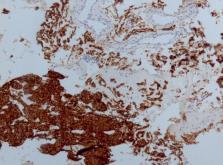
          Le méningiome chordoïde est inscrit selon la dernière classification des tumeurs cérébrales en classe II des méningiomes avec les méningiomes à grandes cellules. C'est une tumeur rare souvent accompagnée de symptômes systémiques décrites par Castelman. Nous présentons un cas de méningiome chordoïde découverte fortuitement chez une patiente de 45 ans suite à un accident de la voie publique, la tomodensitométrie cérébrale réalisée montre une lésion ptérionale gauche intra-diploïque isodense avec lyse osseuse, qui se rehausse de façon homogène après injection de produit de contraste, imagerie par résonnance magnétique montre une lésion hypointense en T1 et spontanément hyperintense en T2/FLAIR, et se rehausse très fortement après injection de gadolinium. Une résection complète de la tumeur a été réalisée. Le diagnostic histologique était de méningiome chordoïde en se basant sur l´étude immuno-histochimique.

          Translated abstract

          Chordoid meningioma is classified in the latest classification of brain tumours as a grade II meningioma, along with large cell meningiomas. It is a rare tumour often associated with systemic symptoms, as described by Castelman. He described a case of chordoid meningioma discovered incidentally in a 45-year-old female patient following road accident. Brain CT scan showed left isodense intradiploid pterional lesion with bone lysis, which was homogeneously enhanced after injection of contrast medium. Magnetic resonance imaging showed T1-hypointense and spontaneously T2 FLAIR hyperintense lesion, which was very strongly enhanced after injection of Gadolinium. Complete resection of the tumour was performed. Histological and immunohistochemical examination showed chordoid meningioma.

          Most cited references10

          • Record: found
          • Abstract: found
          • Article: not found

          The 2021 WHO Classification of Tumors of the Central Nervous System: a summary

          The fifth edition of the WHO Classification of Tumors of the Central Nervous System (CNS), published in 2021, is the sixth version of the international standard for the classification of brain and spinal cord tumors. Building on the 2016 updated fourth edition and the work of the Consortium to Inform Molecular and Practical Approaches to CNS Tumor Taxonomy, the 2021 fifth edition introduces major changes that advance the role of molecular diagnostics in CNS tumor classification. At the same time, it remains wedded to other established approaches to tumor diagnosis such as histology and immunohistochemistry. In doing so, the fifth edition establishes some different approaches to both CNS tumor nomenclature and grading and it emphasizes the importance of integrated diagnoses and layered reports. New tumor types and subtypes are introduced, some based on novel diagnostic technologies such as DNA methylome profiling. The present review summarizes the major general changes in the 2021 fifth edition classification and the specific changes in each taxonomic category. It is hoped that this summary provides an overview to facilitate more in-depth exploration of the entire fifth edition of the WHO Classification of Tumors of the Central Nervous System.
            Bookmark
            • Record: found
            • Abstract: not found
            • Article: not found

            CASE records of the Massachusetts General Hospital Weekly Clinicopathological Exercises: Case 40011.

              Bookmark
              • Record: found
              • Abstract: found
              • Article: not found

              Chordoid meningioma: a clinicopathologic study of 42 cases.

              The term chordoid meningiomas was first used by Kepes et al. in 1987 to describe a meningeal tumor in young patients associated with microcytic anemia and/of dysgammaglobulinemia. Such tumors were composed of spindle or epithelioid cells disposed in chordoma-like clusters and cords in a myxoid matrix and often featured a prominent lymphoplasmacellular infiltrate. Our study includes 42 chordoid meningiomas that represented 0.5% of all meningiomas operated at Mayo Clinic during the interval 1975 to 1997. The male to female ratio was 1:1 and the age range was 12 to 77 years (mean, 47.4 yrs). Only two (5.2%) occurred in children. The majority (88%) were large and supratentorial. No manifestation of systemic disease was noted. Chordoid elements comprised 10% to 100% of the tumors: 34 (81%) were more than 50% chordoid. Thirty-seven tumors (88%) were classified as typical and five as atypical. Lymphoplasmacytic infiltrates varied, being moderate in 10 cases (23.8%), mild in 15 (35.7%), and absent in 17 (40.5%). In 14 (42%) of the 33 cases with available follow up, one or more recurrences were noted. All but one recurrent tumor had been subtotally resected. In 86% of recurrent tumors, the primary lesion was more than 50% chordoid in pattern and contained little or no inflammatory infiltrate. In our experience, chordoid meningiomas are primarily tumors of adults, lack sex predilection, are unassociated with systemic manifestations, and uniformly recur when subtotally excised.
                Bookmark

                Author and article information

                Journal
                Pan Afr Med J
                Pan Afr Med J
                PAMJ
                The Pan African Medical Journal
                The African Field Epidemiology Network
                1937-8688
                13 April 2023
                2023
                : 44
                : 174
                Affiliations
                [1 ]Service de Neurochirurgie, Hôpital Pasteur, Colmar, France
                Author notes
                [& ] Corresponding author: Khalid Skounti, Service de Neurochirurgie, Hôpital Pasteur, Colmar, France. k.skounti@ 123456hotmail.com
                Article
                PAMJ-44-174
                10.11604/pamj.2023.44.174.7539
                10349621
                142ea101-a04a-4e4c-9afd-ed9559f505b4
                Copyright: Khalid Skounti et al.

                The Pan African Medical Journal (ISSN: 1937-8688). This is an Open Access article distributed under the terms of the Creative Commons Attribution International 4.0 License ( https://creativecommons.org/licenses/by/4.0/), which permits unrestricted use, distribution, and reproduction in any medium, provided the original work is properly cited.

                History
                : 20 July 2015
                : 25 November 2015
                Categories
                Case Report

                Medicine
                méningiome chordoïde,maladie de castelman,mib 1,cas clinique,chordoid meningioma,castelman’s disease,case report

                Comments

                Comment on this article