15
views
0
recommends
+1 Recommend
0 collections
    0
    shares
      • Record: found
      • Abstract: found
      • Article: found
      Is Open Access

      Locoregional extension patterns of nasopharyngeal carcinoma and suggestions for clinical target volume delineation

      research-article

      Read this article at

      Bookmark
          There is no author summary for this article yet. Authors can add summaries to their articles on ScienceOpen to make them more accessible to a non-specialist audience.

          Abstract

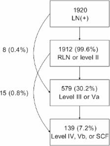
          Clinical target volume (CTV) delineation is crucial for tumor control and normal tissue protection. This study aimed to define the locoregional extension patterns of nasopharyngeal carcinoma (NPC) and to improve CTV delineation. Magnetic resonance imaging scans of 2366 newly diagnosed NPC patients were reviewed. According to incidence rates of tumor invasion, the anatomic sites surrounding the nasopharynx were classified into high-risk (>30%), medium-risk (5%–30%), and low-risk (<5%) groups. The lymph node (LN) level was determined according to the Radiation Therapy Oncology Group guidelines, which were further categorized into the upper neck (retropharyngeal region and level II), middle neck (levels III and Va), and lower neck (levels IV and Vb and the supraclavicular fossa). The high-risk anatomic sites were adjacent to the nasopharynx, whereas those at medium- or low-risk were separated from the nasopharynx. If the high-risk anatomic sites were involved, the rates of tumor invasion into the adjacent medium-risk sites increased; if not, the rates were significantly lower ( P < 0.01). Among the 1920 (81.1%) patients with positive LN, the incidence rates of LN metastasis in the upper, middle, and lower neck were 99.6%, 30.2%, and 7.2%, respectively, and skip metastasis happened in only 1.2% of patients. In the 929 patients who had unilateral upper neck involvement, the rates of contralateral middle neck and lower neck involvement were 1.8% and 0.4%, respectively. Thus, local disease spreads stepwise from proximal sites to distal sites, and LN metastasis spreads from the upper neck to the lower neck. Individualized CTV delineation for NPC may be feasible.

          Related collections

          Most cited references29

          • Record: found
          • Abstract: found
          • Article: not found

          How does intensity-modulated radiotherapy versus conventional two-dimensional radiotherapy influence the treatment results in nasopharyngeal carcinoma patients?

          To compare the results of intensity-modulated radiotherapy (IMRT) with those of two-dimensional conventional radiotherapy (2D-CRT) in the treatment of patients with nasopharyngeal carcinoma (NPC). A retrospective review of data from 1,276 patients with biopsy-proven, nonmetastatic NPC was performed. All patients had undergone magnetic resonance imaging and were staged according to the sixth edition of the American Joint Committee on Cancer staging criteria. Radiotherapy was the primary treatment for all patients. Of the 1,276 patients, 512 were treated with IMRT and 764 with 2D-CRT. The 5-year actuarial local relapse-free survival (LRFS), the nodal relapse-free survival (NRFS), the distant metastasis-free survival (DMFS), and the disease-free survival (DFS) rates were 92.7%, 97.0%, 84.0%, and 75.9%, respectively, for the IMRT group, and 86.8%, 95.5%, 82.6%, and 71.4%, respectively, for the 2D-CRT group. In stage T1 patients, improvement of LRFS in the IMRT group was even significantly higher than in the 2D-CRT group (100% vs. 94.4%; p = 0.016). A trend of improvement of DFS was observed in the IMRT group compared with the 2D-CRT group but without reaching statistical significance. NRFS and DMFS rates were similar in the two groups. A greater improvement of treatment results with IMRT than with 2D-CRT was demonstrated primarily by achieving a higher local tumor control rate in NPC patients, especially in the early T stage patients. The goal of better control of both local failure in advanced, nonmetastatic NPC patients and of distant failure should be addressed in future studies. Copyright © 2011 Elsevier Inc. All rights reserved.
            Bookmark
            • Record: found
            • Abstract: found
            • Article: not found

            Intensity-modulated radiotherapy in the treatment of nasopharyngeal carcinoma: an update of the UCSF experience.

            To update our experience with intensity-modulated radiotherapy (IMRT) in the treatment of nasopharyngeal carcinoma (NPC). Between April 1995 and October 2000, 67 patients underwent IMRT for NPC at the University of California-San Francisco (UCSF). There were 20 females and 47 males, with a mean age of 49 (range 17-82). The disease was Stage I in 8 (12%), Stage II in 12 (18%), Stage III in 22 (33%), and Stage IV in 25 (37%). IMRT was delivered using three different techniques: 1) manually cut partial transmission blocks, 2) computer-controlled auto-sequencing segmental multileaf collimator (SMLC), and 3) sequential tomotherapy using a dynamic multivane intensity modulating collimator (MIMiC). Fifty patients received concomitant cisplatinum and adjuvant cisplatinum and 5-FU chemotherapy according to the Intergroup 0099 trial. Twenty-six patients had fractionated high-dose-rate intracavitary brachytherapy boost and 1 patient had gamma knife radiosurgery boost after external beam radiotherapy. The prescribed dose was 65-70 Gy to the gross tumor volume (GTV) and positive neck nodes, 60 Gy to the clinical target volume (CTV), 50-60 Gy to the clinically negative neck, and 5-7 Gy in 2 fractions for the intracavitary brachytherapy boost. Acute and late normal tissue effects were graded according to the Radiation Therapy Oncology Group (RTOG) radiation morbidity scoring criteria. The local progression-free, local-regional progression-free, distant metastasis-free rates, and the overall survival were calculated using the Kaplan-Meier method. With a median follow-up of 31 months (range 7 to 72 months), there has been one local recurrence at the primary site. One patient failed in the neck. Seventeen patients developed distant metastases; 5 of these patients have died. The 4-year estimates of local progression-free, local-regional progression-free, and distant metastases-free rates were 97%, 98%, and 66% respectively. The 4-year estimate of overall survival was 88%. The worst acute toxicity documented was as follows: Grade 1 or 2 in 51 patients, Grade 3 in 15 patients, and Grade 4 in 1 patient. The worst late toxicity was Grade 1 in 20 patients, Grade 2 in 15 patients, Grade 3 in 7 patients, and Grade 4 in 1 patient. At 3 months after IMRT, 64% of the patients had Grade 2, 28% had Grade 1, and 8% had Grade 0 xerostomia. Xerostomia decreased with time. At 24 months, only one of the 41 evaluable patients had Grade 2, 32% had Grade 1, and 66% had Grade 0 or no xerostomia. Analysis of the dose-volume histograms (DVHs) showed that the average maximum, mean, and minimum dose delivered were 79.3 Gy, 74.5 Gy, and 49.4 Gy to the GTV, and 78.9 Gy, 68.7 Gy, and 36.8 Gy to the CTV. An average of only 3% of the GTV and 3% of the CTV received less than 95% of the prescribed dose. Excellent local-regional control for NPC was achieved with IMRT. IMRT provided excellent tumor target coverage and allowed the delivery of a high dose to the target with significant sparing of the salivary glands and other nearby critical normal tissues.
              Bookmark
              • Record: found
              • Abstract: found
              • Article: not found

              Treatment of nasopharyngeal carcinoma with intensity-modulated radiotherapy: the Hong Kong experience.

              To evaluate the efficacy of using intensity-modulated radiotherapy (IMRT) in the primary treatment of nasopharyngeal carcinoma (NPC), including the role of dose escalation above 66 Gy level. Between July 2000 and September 2002, 63 newly diagnosed NPC patients were treated with IMRT. The disease was Stage I in 9 (14%), Stage II in 18 (29%), Stage III in 22 (35%), and Stage IV in 14 (22%). The prescribed dose was 66 Gy to the gross tumor volume (GTV) and positive neck nodes, 60 Gy to the planning target volume (PTV), and 54-60 Gy to the clinically negative neck. All 20 (100%) patients with T1-2a tumors received intracavitary brachytherapy (ICB) boost, and 15/42 (36%) patients with T2b-T4 tumors received conformal boost (8 Gy/4 fractions). Nineteen patients with advanced stage disease also received either neoadjuvant or concurrent chemotherapy. Acute and late normal tissue effects were graded according to the Radiation Therapy Oncology Group (RTOG) radiation morbidity scoring criteria. Local relapse-free survival (LRFS), nodal relapse-free survival (NRFS), distant metastasis-free survival (DMFS), and overall survival (OS) were estimated using the Kaplan-Meier method. With a median follow-up of 29 months (range 8-45 months), 4 patients developed local in-field failure, 1 patient developed regional relapse, and 13 patients developed distant metastases. All 4 patients with local failure had either T3 or T4 disease before primary treatment and did not have ICB or conformal boost. The 3-year actuarial LRFS, NRFS, DMFS, and OS were 92%, 98%, 79%, and 90%, respectively. Multivariate analysis showed that dose escalation above 66 Gy was significantly associated with better PFS and DMFS, whereas GTV size was a significant adverse factor for OS. The worst acute mucositis was Grade 1 or 2 in 36 (59%), and Grade 3 in 25 (41%) patients. Acute dysphagia requiring tube feeding occurred in 5 (8%) patients. The proportion of patients with Grade 2-3 xerostomia was 57% at 3 months, and 23% at 2 years after IMRT. Within the subset of patients with a mean parotid dose of <31 Gy, the proportions with Grade 2-3 xerostomia were 30% and 17% at 3 months and 2 years, respectively. Our experience of using IMRT in the primary treatment of NPC showed a very high rate of locoregional control and favorable toxicity profile. Furthermore, we found that dose escalation above 66 Gy of IMRT-based therapy was a significant determinant of progression-free survival and distant metastasis-free survival for advanced T-stage tumors. Distant metastases represent the predominant mode of treatment failure.
                Bookmark

                Author and article information

                Journal
                Chin J Cancer
                Chin J Cancer
                CJC
                Chinese Journal of Cancer
                Sun Yat-sen University Cancer Center
                1000-467X
                1944-446X
                December 2012
                : 31
                : 12
                : 579-587
                Affiliations
                [1 ]State Key Laboratory of Oncology in South China, Guangzhou, Guangdong 510060, P. R. China;
                [2 ]Department of Radiation Oncology,
                [3 ]Imaging Diagnosis and Interventional Center, Sun Yat-sen University Cancer Center, Guangzhou, Guangdong 510060, P. R. China.
                Author notes
                Corresponding Author: Jun Ma, Department of Radiation Oncology, Sun Yat-sen University Cancer Center, 651 Dongfeng Road East, Guangzhou, Guangdong 510060, P. R. China. Tel: +86-20-87343469; Email: majun@ 123456sysucc.org.cn .
                Article
                cjc-31-12-579
                10.5732/cjc.012.10095
                3777458
                22854064
                15661c54-41d7-49f1-8790-d8b6e4620804
                Chinese Journal of Cancer

                This is an open-access article distributed under the terms of the Creative Commons Attribution-NonCommercial-ShareAlike 3.0 Unported License, which allows readers to alter, transform, or build upon the article and then distribute the resulting work under the same or similar license to this one. The work must be attributed back to the original author and commercial use is not permitted without specific permission.

                History
                : 15 April 2012
                : 11 June 2012
                : 4 July 2012
                Categories
                Original Article

                nasopharyngeal carcinoma,magnetic resonance imaging,local extension,lymph node spread,clinical target volume

                Comments

                Comment on this article