46
views
0
recommends
+1 Recommend
0 collections
    0
    shares
      • Record: found
      • Abstract: found
      • Article: not found

      The Role of Mir-148a in Cancer

      review-article

      Read this article at

      ScienceOpenPublisherPMC
      Bookmark
          There is no author summary for this article yet. Authors can add summaries to their articles on ScienceOpen to make them more accessible to a non-specialist audience.

          Abstract

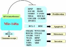
          MicroRNAs (miRNAs) are highly conserved noncoding RNAs of about 19-25 nucleotides. Through specifically pairing with complementary sites in 3' untranslated regions (UTRs) of target mRNAs, they mediate post-transcriptional silencing. MicroRNAs have been implicated in many physiological processes including proliferation, differentiation, development, apoptosis, and metabolism. In recent years many studies have revealed that the aberrant expression of miRNA is closely related to oncogenesis and is now an intense field of study. Mir-148a is aberrantly expressed in various cancers and has been identified as an oncogenic or tumor suppressor with crucial roles in the molecular mechanisms of oncogenesis. In this review, we have summarized the role of mir-148a in the oncogenic pathways of gastric, liver, breast and urogenital cancers, and in neurogliocytoma oncogenesis. Studying the functional role of mir-148a is crucial in discovering novel tumor molecular markers and identifying potential therapeutic targets.

          Related collections

          Most cited references80

          • Record: found
          • Abstract: found
          • Article: not found

          MicroRNAs in human cancer.

          Mature microRNAs (miRNAs) are single-stranded RNA molecules of 20-23-nucleotide (nt) length that control gene expression in many cellular processes. These molecules typically reduce the translation and stability of mRNAs, including those of genes that mediate processes in tumorigenesis, such as inflammation, cell cycle regulation, stress response, differentiation, apoptosis, and invasion. miRNA targeting is initiated through specific base-pairing interactions between the 5' end ("seed" region) of the miRNA and sites within coding and untranslated regions (UTRs) of mRNAs; target sites in the 3' UTR lead to more effective mRNA destabilization. Since miRNAs frequently target hundreds of mRNAs, miRNA regulatory pathways are complex. To provide a critical overview of miRNA dysregulation in cancer, we first discuss the methods currently available for studying the role of miRNAs in cancer and then review miRNA genomic organization, biogenesis, and mechanism of target recognition, examining how these processes are altered in tumorigenesis. Given the critical role miRNAs play in tumorigenesis processes and their disease specific expression, they hold potential as therapeutic targets and novel biomarkers.
            Bookmark
            • Record: found
            • Abstract: found
            • Article: not found

            MicroRNA expression alterations are linked to tumorigenesis and non-neoplastic processes in pancreatic ductal adenocarcinoma.

            Pancreatic ductal adenocarcinoma (PDAC) is known for its very poor overall prognosis. Accurate early diagnosis and new therapeutic modalities are therefore urgently needed. We used 377 feature microRNA (miRNA) arrays to investigate miRNA expression in normal pancreas, chronic pancreatitis, and PDAC tissues as well as PDAC-derived cell lines. A pancreatic miRNome was established comparing the data from normal pancreas with a reference set of 33 human tissues. The expression of miR-216 and -217 and lack of expression of miR-133a were identified as characteristic of pancreas tissue. Unsupervised clustering showed that the three pancreatic tissues types can be classified according to their respective miRNA expression profiles. We identified 26 miRNAs most prominently misregulated in PDAC and a relative quantitative reverse transcriptase-polymerase chain reaction index using only miR-217 and -196a was found to discriminate normal pancreas, chronic pancreatitis and cancerous tissues, establishing a potential utility for miRNAs in diagnostic procedures. Lastly, comparing differentially expressed genes from PDAC with predicted miRNA target genes for the top 26 miRNAs, we identified potential novel links between aberrant miRNA expression and known target genes relevant to PDAC biology. Our data provides novel insights into the miRNA-driven pathophysiological mechanisms involved in PDAC development and offers new candidate targets to be exploited both for diagnostic and therapeutic strategies.
              Bookmark
              • Record: found
              • Abstract: found
              • Article: not found

              A targeting sequence directs DNA methyltransferase to sites of DNA replication in mammalian nuclei.

              Tissue-specific patterns of methylated deoxycytidine residues in the mammalian genome are preserved by postreplicative methylation of newly synthesized DNA. DNA methyltransferase (MTase) is here shown to associate with replication foci during S phase but to display a diffuse nucleoplasmic distribution in non-S phase cells. Analysis of DNA MTase-beta-galactosidase fusion proteins has shown that association with replication foci is mediated by a novel targeting sequence located near the N-terminus of DNA MTase. This sequence has the properties expected of a targeting sequence in that it is not required for enzymatic activity, prevents proper targeting when deleted, and, when fused to beta-galactosidase, causes the fusion protein to associate with replication foci in a cell cycle-dependent manner.
                Bookmark

                Author and article information

                Journal
                J Cancer
                J Cancer
                jca
                Journal of Cancer
                Ivyspring International Publisher (Sydney )
                1837-9664
                2016
                21 June 2016
                : 7
                : 10
                : 1233-1241
                Affiliations
                1. Department of Pathology and Pathophysiology, Hunan Normal University Medical School, Changsha 410013, Hunan, China;
                2. Department of Statistics and Epidemiology, Public Health School, Central South University, Changsha 410078, Hunan, China.
                Author notes
                ✉ Corresponding authors: Xiaoning Peng, MD. Department of Pathology and Pathophysiology, Hunan Normal University Medical School, Changsha 410013, Hunan, China. Tel: +86-13786169507 Fax: +86-21-64085875 E-mail: pxiaoning@ 123456hunnu.edu.cn or Xiaomin Zeng, MS. Department of Statistics and Epidemiology, Public Health School, Central South University, Changsha, Hunan410078, China. Tel: +86-13467630366 Fax: +86-21-64085875 E-mail: zxiaomin@ 123456csu.edu.cn .

                Conflict of Interests: All authors declare that they have no any conflict of interests.

                Article
                jcav07p1233
                10.7150/jca.14616
                4934031
                27390598
                16f38d48-3727-4ad3-a6cd-d978d5d9bc4e
                © Ivyspring International Publisher. Reproduction is permitted for personal, noncommercial use, provided that the article is in whole, unmodified, and properly cited. See http://ivyspring.com/terms for terms and conditions.
                History
                : 7 December 2015
                : 7 May 2016
                Categories
                Review

                Oncology & Radiotherapy
                mirna,mir-148a,cancer,oncogene,tumor suppressor gene,biological function.
                Oncology & Radiotherapy
                mirna, mir-148a, cancer, oncogene, tumor suppressor gene, biological function.

                Comments

                Comment on this article