13
views
0
recommends
+1 Recommend
0 collections
    0
    shares
      • Record: found
      • Abstract: found
      • Article: found
      Is Open Access

      Quantitation of the cellular content of saliva and buccal swab samples

      research-article

      Read this article at

      Bookmark
          There is no author summary for this article yet. Authors can add summaries to their articles on ScienceOpen to make them more accessible to a non-specialist audience.

          Abstract

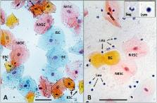
          Buccal swabs and saliva are the two most common oral sampling methods used for medical research. Often, these samples are used interchangeably, despite previous evidence that both contain buccal cells and blood leukocytes in different proportions. For some research, such as epigenetic studies, the cell types contributing to the analysis are highly relevant. We collected such samples from twelve children and twenty adults and, using Papanicolaou staining, measured the proportions of epithelial cells and leukocytes through microscopy. To our knowledge, no studies have compared cellular heterogeneity in buccal swab and saliva samples from adults and children. We confirmed that buccal swabs contained a higher proportion of epithelial cells than saliva and that children have a greater proportion of such cells in saliva compared to adults. At this level of resolution, buccal swabs and saliva contained similar epithelial cell subtypes. Gingivitis in children was associated with a higher proportion of leukocytes in saliva samples but not in buccal swabs. Compared to more detailed and costly methods such as flow cytometry or deconvolution methods used in epigenomic analysis, the procedure described here can serve as a simple and low-cost method to characterize buccal and saliva samples. Microscopy provides a low-cost tool to alert researchers to the presence of oral inflammation which may affect a subset of their samples. This knowledge might be highly relevant to their specific research questions, may assist with sample selection and thus might be crucial information despite the ability of data deconvolution methods to correct for cellular heterogeneity.

          Related collections

          Most cited references22

          • Record: found
          • Abstract: found
          • Article: not found

          DNA extracted from saliva for methylation studies of psychiatric traits: evidence tissue specificity and relatedness to brain.

          DNA methylation has become increasingly recognized in the etiology of psychiatric disorders. Because brain tissue is not accessible in living humans, epigenetic studies are most often conducted in blood. Saliva is often collected for genotyping studies but is rarely used to examine DNA methylation because the proportion of epithelial cells and leukocytes varies extensively between individuals. The goal of this study was to evaluate whether saliva DNA is informative for studies of psychiatric disorders. DNA methylation (HumanMethylation450 BeadChip) was assessed in saliva and blood samples from 64 adult African Americans. Analyses were conducted using linear regression adjusted for appropriate covariates, including estimated cellular proportions. DNA methylation from brain tissues (cerebellum, frontal cortex, entorhinal cortex, and superior temporal gyrus) was obtained from a publically available dataset. Saliva and blood methylation was clearly distinguishable though there was positive correlation overall. There was little correlation in CpG sites within relevant candidate genes. Correlated CpG sites were more likely to occur in areas of low CpG density (i.e., CpG shores and open seas). There was more variability in CpG sites from saliva than blood, which may reflect its heterogeneity. Finally, DNA methylation in saliva appeared more similar to patterns from each of the brain regions examined overall than methylation in blood. Thus, this study provides a framework for using DNA methylation from saliva and suggests that DNA methylation of saliva may offer distinct opportunities for epidemiological and longitudinal studies of psychiatric traits.
            Bookmark
            • Record: found
            • Abstract: found
            • Article: not found

            Buccal micronucleus cytome assay.

            The Buccal Micronucleus Cytome (BMCyt) assay is a minimally invasive method for studying DNA damage, chromosomal instability, cell death and the regenerative potential of human buccal mucosal tissue. This method is increasingly used in molecular epidemiological studies for investigating the impact of nutrition, lifestyle factors, genotoxin exposure and genotype on DNA damage, chromosome malsegregation and cell death. The biomarkers measured in this assay have been associated with increased risk of accelerated ageing, cancer and neurodegenerative diseases. This protocol describes one of the current established methods for buccal cell collection using a small-headed toothbrush, the generation of a single-cell suspension, slide preparation using cytocentrifugation, fixation and staining using Feulgen and Light Green for both bright field and fluorescence microscopic analysis. The scoring criteria for micronuclei and other nuclear anomalies are also described in detail. The protocol in its current form takes approximately 4 h to complete from the time of buccal cell collection to the generation of stained slides for microscopic analysis.
              Bookmark
              • Record: found
              • Abstract: found
              • Article: found
              Is Open Access

              Reference-free deconvolution of DNA methylation data and mediation by cell composition effects

              Background Recent interest in reference-free deconvolution of DNA methylation data has led to several supervised methods, but these methods do not easily permit the interpretation of underlying cell types. Results We propose a simple method for reference-free deconvolution that provides both proportions of putative cell types defined by their underlying methylomes, the number of these constituent cell types, as well as a method for evaluating the extent to which the underlying methylomes reflect specific types of cells. We demonstrate these methods in an analysis of 23 Infinium data sets from 13 distinct data collection efforts; these empirical evaluations show that our algorithm can reasonably estimate the number of constituent types, return cell proportion estimates that demonstrate anticipated associations with underlying phenotypic data; and methylomes that reflect the underlying biology of constituent cell types. Conclusions Our methodology permits an explicit quantitation of the mediation of phenotypic associations with DNA methylation by cell composition effects. Although more work is needed to investigate functional information related to estimated methylomes, our proposed method provides a novel and useful foundation for conducting DNA methylation studies on heterogeneous tissues lacking reference data. Electronic supplementary material The online version of this article (doi:10.1186/s12859-016-1140-4) contains supplementary material, which is available to authorized users.
                Bookmark

                Author and article information

                Contributors
                jeffrey.craig@deakin.edu.au
                Journal
                Sci Rep
                Sci Rep
                Scientific Reports
                Nature Publishing Group UK (London )
                2045-2322
                2 May 2018
                2 May 2018
                2018
                : 8
                : 6944
                Affiliations
                [1 ]The Royal Women’s Hospital, Parkville, Victoria, Australia
                [2 ]ISNI 0000 0000 9442 535X, GRID grid.1058.c, Murdoch Children’s Research Institute, Parkville, ; Victoria, Australia
                [3 ]ISNI 0000 0001 2179 088X, GRID grid.1008.9, Department of Obstetrics and Gynaecology, , University of Melbourne, Parkville, ; Victoria, Australia
                [4 ]ISNI 0000 0001 2179 088X, GRID grid.1008.9, Department of Pathology, , University of Melbourne, Parkville, ; Victoria, Australia
                [5 ]ISNI 0000 0001 2179 088X, GRID grid.1008.9, Department of Paediatrics, , University of Melbourne, Royal Children’s Hospital, ; Victoria, Australia
                [6 ]ISNI 0000 0001 0526 7079, GRID grid.1021.2, Centre for Molecular and Medical Research, , School of Medicine, Deakin University, Geelong, ; Victoria, Australia
                Author information
                http://orcid.org/0000-0001-7577-6004
                Article
                25311
                10.1038/s41598-018-25311-0
                5932057
                29720614
                17bcf355-f421-409c-87fe-f9c96dcf58d9
                © The Author(s) 2018

                Open Access This article is licensed under a Creative Commons Attribution 4.0 International License, which permits use, sharing, adaptation, distribution and reproduction in any medium or format, as long as you give appropriate credit to the original author(s) and the source, provide a link to the Creative Commons license, and indicate if changes were made. The images or other third party material in this article are included in the article’s Creative Commons license, unless indicated otherwise in a credit line to the material. If material is not included in the article’s Creative Commons license and your intended use is not permitted by statutory regulation or exceeds the permitted use, you will need to obtain permission directly from the copyright holder. To view a copy of this license, visit http://creativecommons.org/licenses/by/4.0/.

                History
                : 11 October 2017
                : 17 April 2018
                Categories
                Article
                Custom metadata
                © The Author(s) 2018

                Uncategorized
                Uncategorized

                Comments

                Comment on this article