54
views
0
recommends
+1 Recommend
1 collections
    0
    shares
      Are you tired of sifting through news that doesn't interest you?
      Personalize your Karger newsletter today and get only the news that matters to you!

      Sign up

      • Record: found
      • Abstract: found
      • Article: found
      Is Open Access

      Ectopic Cilia: A Histopathological Study

      case-report

      Read this article at

      ScienceOpenPublisherPMC
      Bookmark
          There is no author summary for this article yet. Authors can add summaries to their articles on ScienceOpen to make them more accessible to a non-specialist audience.

          Abstract

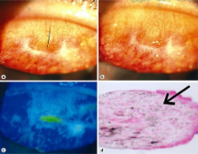
          Cilia are normally found at the eyelid margin, while ectopic cilia are one or more lash follicles appearing in an abnormal position within the eyelid. We herein report two cases of cilia located in the palpebral conjunctiva. A 31-year-old female and a 46-year-old male presented with ectopic cilia in the superior palpebral conjunctiva. Histopathological study of the excised ectopic cilia and related lesions showed the cilia-related lesion to be located in the epithelial pit that contains goblet cells, which is consistent with the crypts of Henle. The hair follicle was surrounded by granulation tissue, while a dermal papilla and a hair matrix, which are known to produce hair follicles, did not exist in the excised tissue. While anterior ectopic cilia are congenital, ectopic cilia in the palpebral conjunctiva may be acquired, and these aberrant cilia are associated with crypts of Henle and chronic inflammation.

          Related collections

          Most cited references4

          • Record: found
          • Abstract: found
          • Article: not found

          ''Ectopic eyelashes'' (ectopic cilia) in a 2-year-old girl: brief report and discussion of possible embryologic origin.

          Cilia, or eyelashes, are unique hair follicles normally found at the eyelid margin. The spectrum of cilial anomalies includes cilial row duplication, agenesis, and ectopic placement. Ectopic cilia are the most rare of cilial anomalies. We report a case of a 2-and-a-half-year-old girl with ectopic cilia of the anterior tarsal plate, an extremely rare, congenital anomaly that is most often not associated with other findings and likely results from an event during embryogenesis.
            Bookmark
            • Record: found
            • Abstract: found
            • Article: not found

            Ectopic cilia in a 14-year-old boy.

            Ectopic cilia is a very rare condition in human beings, usually presented as a tuft of densely grouped hair on the lateral quadrant of the upper eyelid or conjunctival surface of the eyelid. The embryologic origin of this anomaly is unknown, and few cases have been recorded in literature. In most cases, it causes no apparent medical morbidity but is treated by surgical excision because of its cosmetic aspect. We report a 14-year-old boy with ectopic cilia with approximately 20 hairs occurring as single and grouped hairs pattern emerging from anterior surface of eyelid. Positive history of the same anomaly in his paternal grandfather demonstrates evidence of an inherited genetic disorder. © 2011 Wiley Periodicals, Inc.
              Bookmark
              • Record: found
              • Abstract: found
              • Article: not found

              A case of ectopic cilia.

              Ectopic cilia are the most rare of cilial anomalies. We report a 27-year-old man who presents with tarsal conjunctival symptomatic ectopic cilia. On slit-lamp examination eversion of the lower eyelid demonstrated that several black-white cilia were protruding from the conjunctiva.
                Bookmark

                Author and article information

                Journal
                Case Rep Dermatol
                Case Rep Dermatol
                CDE
                Case Reports in Dermatology
                S. Karger AG (Allschwilerstrasse 10, P.O. Box · Postfach · Case postale, CH–4009, Basel, Switzerland · Schweiz · Suisse, Phone: +41 61 306 11 11, Fax: +41 61 306 12 34, karger@karger.ch )
                1662-6567
                Jan-Apr 2012
                10 February 2012
                10 February 2012
                : 4
                : 1
                : 37-40
                Affiliations
                [1] aDepartment of Ophthalmology, Hokkaido University Graduate School of Medicine, Sapporo, Japan
                [2] bDepartment of Dermatology, Hokkaido University Graduate School of Medicine, Sapporo, Japan
                [3] cDepartment of Ohashi Eye Center, Sapporo, Japan
                Author notes
                *Satoru Kase, Department of Ophthalmology, Hokkaido University Graduate School of Medicine, Nishi 7, Kita 15, Kita-ku, Sapporo 060-8638 (Japan), Tel. +81 11 706 5944, E-Mail kaseron@ 123456med.hokudai.ac.jp
                Article
                cde-0004-0037
                10.1159/000336887
                3355649
                22619655
                17e47536-dbd1-4565-8072-4f49b4ecf4f2
                Copyright © 2012 by S. Karger AG, Basel

                This is an Open Access article distributed under the terms of the Creative Commons Attribution-Noncommercial-No-Derivative-Works License ( http://creativecommons.org/licenses/by-nc-nd/3.0/). Users may download, print and share this work on the Internet for noncommercial purposes only, provided the original work is properly cited, and a link to the original work on http://www.karger.com and the terms of this license are included in any shared versions.

                History
                Page count
                Figures: 2, References: 4, Pages: 4
                Categories
                Published: February, 2012

                Dermatology
                crypts of henle,histopathology,ectopic cilia
                Dermatology
                crypts of henle, histopathology, ectopic cilia

                Comments

                Comment on this article