331
views
0
recommends
+1 Recommend
0 collections
    16
    shares
      • Record: found
      • Abstract: found
      • Article: found
      Is Open Access

      Assessment of the Spatial QRS-T Angle by Vectorcardiography: Current Data and Perspectives

      review-article

      Read this article at
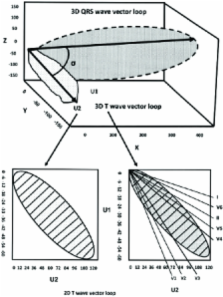
      Bookmark
          There is no author summary for this article yet. Authors can add summaries to their articles on ScienceOpen to make them more accessible to a non-specialist audience.

          Abstract

          The concept of the ventricular gradient (VG) was conceived in the 1930s and its calculation yielded information that was not otherwise obtainable. The VG was not utilized by clinicians at large because it was not easy to understand and its computation time-consuming. Spatial vectorcardiography is based on the concept of the VG. Its current major clinical use is to identify primary [heterogeneity of ventricular action potential (VAP) morphology] in the presence of secondary [heterogeneity in ventricular depolarization instants] T-wave abnormalities in an ECG. Nowadays, the calculation of the spatial VG can be computed on the basis of a regular routine ECG and contributes to localization of arrhythmogenic areas in the heart by assessing overall and local VAP duration heterogeneity. Recent population-based studies suggest that the spatial VG is a dominant ECG predictor of future cardiovascular events and death and it is superior to more conventional ECG parameters. Its assessment warrants consideration for intensified primary and secondary prevention efforts and can be included in everyday clinical practice. This review addresses the nature and diagnostic potential of the spatial VG. The main focus is the role of the spatial VG in ECG assessment of dispersion of repolarization, a key factor in arrhythmogeneity.

          Related collections

          Most cited references102

          • Record: found
          • Abstract: found
          • Article: not found

          National study of physician awareness and adherence to cardiovascular disease prevention guidelines.

          Few data have evaluated physician adherence to cardiovascular disease (CVD) prevention guidelines according to physician specialty or patient characteristics, particularly gender. An online study of 500 randomly selected physicians (300 primary care physicians, 100 obstetricians/gynecologists, and 100 cardiologists) used a standardized questionnaire to assess awareness of, adoption of, and barriers to national CVD prevention guidelines by specialty. An experimental case study design tested physician accuracy and determinants of CVD risk level assignment and application of guidelines among high-, intermediate-, or low-risk patients. Intermediate-risk women, as assessed by the Framingham risk score, were significantly more likely to be assigned to a lower-risk category by primary care physicians than men with identical risk profiles (P<0.0001), and trends were similar for obstetricians/gynecologists and cardiologists. Assignment of risk level significantly predicted recommendations for lifestyle and preventive pharmacotherapy. After adjustment for risk assignment, the impact of patient gender on preventive care was not significant except for less aspirin (P<0.01) and more weight management recommended (P<0.04) for intermediate-risk women. Physicians did not rate themselves as very effective in their ability to help patients prevent CVD. Fewer than 1 in 5 physicians knew that more women than men die each year from CVD. Perception of risk was the primary factor associated with CVD preventive recommendations. Gender disparities in recommendations for preventive therapy were explained largely by the lower perceived risk despite similar calculated risk for women versus men. Educational interventions for physicians are needed to improve the quality of CVD preventive care and lower morbidity and mortality from CVD for men and women.
            Bookmark
            • Record: found
            • Abstract: found
            • Article: not found

            Cycle length dependence of human action potential duration in vivo. Effects of single extrastimuli, sudden sustained rate acceleration and deceleration, and different steady-state frequencies.

            Using a new method for long-term recording of monophasic action potentials from the human heart, we studied in 17 patients the effects on ventricular action potential duration (APD) of three clinically pertinent cycle length perturbations: (1) single extrastimuli, (2) abrupt sustained rate acceleration and deceleration, and (3) different steady-state cycle lengths. Results were: (a) APD after single extrastimuli at progressively longer cycle lengths were related to the extrastimulus cycle length with a biphasic electrical restitution curve which after an initial steep rise and a subsequent transient descent rose again more gradually to a plateau at cycle lengths above 800-1,000 ms. (b) After a sustained step decrease in cycle length, the first APD shortened abruptly while final steady-state adaptation required up to several minutes. The transition between the rapid and slow phase of APD change was characterized by a variable alternans of APD which correlated inversely with the preceding diastolic interval. (c) In the steady state, APD correlated linearly with cycle length, increasing an average of 23 ms per 100 ms cycle length increase (r = 0.995). The divergence between steady-state and non-steady-state APD, and the slowness of steady-state adaptation, are important factors to be considered in clinical electrophysiologic studies and in rate correction algorithms of APD or QT intervals, respectively.
              Bookmark
              • Record: found
              • Abstract: found
              • Article: not found

              Characteristics and possible mechanism of ventricular arrhythmia dependent on the dispersion of action potential durations.

              The arrhythmogenic role of increased dispersion of repolarization (dispersion) was studied in 23 open-chest dogs using six simultaneously recorded monophasic action potentials (MAPs) from the ventricular surface and programmed ventricular premature stimulation (VPS). Increased dispersion was induced by generalized hypothermia (29 degrees C) and regional warm blood (38-43 degrees C) perfusion through a coronary artery branch. Hypothermia and regional warm blood perfusion increased maximum dispersion from 13 +/- 10 to 111 +/- 16 msec (p less than 0.001), predominantly because of the increased MAP duration difference (10 +/- 15 vs 97 +/- 16 msec, p less than 0.001). The maximal difference between activation times was not significantly changed, but the QRS duration increased from 47 +/- 6 to 52 +/- 7 msec (p less than 0.01). Ventricular arrhythmia did not occur spontaneously but was induced by a single VPS in all 23 dogs during hypothermia and regional warm blood perfusion when dispersion reached a critical magnitude. The critical magnitude of dispersion required to induce ventricular arrhythmia was documented in 16 dogs by stepwise increments or decrements of dispersion. In four dogs, an increase in atrial pacing rate of 24 beats/min prevented induction of ventricular arrhythmia by decreasing dispersion from a critical magnitude of 103 +/- 5 msec to a nonarrhythmogenic value of 86 +/- 9 msec (p less than 0.05). In six dogs, we compared the stimulation site-dependent effects of VPS applied in the region with short and long MAPs. In all dogs, ventricular arrhythmia was inducible only by VPS from the region with a short MAP. Premature impulses from this region propagated more slowly than those from the region with a long MAP. Our results show that the large dispersion of repolarization facilitates the development of a conduction delay necessary to induce sustained arrhythmia by an early premature stimulus applied at the site with a short MAP.
                Bookmark

                Author and article information

                Journal
                Curr Cardiol Rev
                CCR
                Current Cardiology Reviews
                Bentham Science Publishers Ltd.
                1573-403X
                1875-6557
                November 2009
                : 5
                : 4
                : 251-262
                Affiliations
                []1st Department of Propaedeutic Medicine, Athens University Medical School, Laiko General Hospital Athens, Greece
                Author notes
                [* ]Address correspondence to this author at the 1st Department of Propaedeutic Medicine, Athens University Medical School, Laiko General Hospital, 33 Lakonias Street, 11523 Athens, Greece; Tel: +30 210 745 6261; Fax: +30 210 746 2640; E-mail: ntentol@ 123456med.uoa.gr
                Article
                CCR-5-251
                10.2174/157340309789317850
                2842956
                21037841
                18d04991-f123-4d19-9b56-7bcde14d347e
                © 2009 Bentham Science Publishers Ltd.

                This is an open access article distributed under the terms of the Creative Commons Attribution License ( http://creativecommons.org/licenses/by/2.5/), which permits unrestrictive use, distribution, and reproduction in any medium, provided the original work is properly cited.

                History
                : 22 August 2008
                : 26 March 2009
                : 31 March 2009
                Categories
                Article

                Cardiovascular Medicine
                action potential duration,spatial qrs-t angle,vectorcardiography,arrhythmogenicity.,spatial ventricular gradient

                Comments

                Comment on this article