3
views
0
recommends
+1 Recommend
0 collections
    0
    shares
      • Record: found
      • Abstract: found
      • Article: found
      Is Open Access

      Thalamic Deep Brain Stimulation for Refractory Atypical Tremor after Encephalitis of Unknown Etiology: A Case Report

      case-report

      Read this article at

      Bookmark
          There is no author summary for this article yet. Authors can add summaries to their articles on ScienceOpen to make them more accessible to a non-specialist audience.

          Abstract

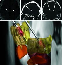
          Tremor associated with encephalitis is usually transient and rarely becomes chronic and refractory. Treatment for such tremor using deep brain stimulation (DBS) has not yet been reported. We report an uncommon case of chronic tremor after encephalitis of unknown etiology and its outcome treated with thalamic DBS. A 47-year-old man presented with a 6-month history of medically refractory tremor after non-infectious and probable autoimmune encephalitis. The patient showed an atypical mixture of resting, postural, kinetic, and intention tremor. The tremor significantly disabled the patient’s activities of daily life (ADL). The patient underwent bilateral thalamic DBS surgery. DBS leads were placed to cross the border between the ventralis oralis posterior (Vop) nucleus and ventralis intermedius (Vim) nucleus of the thalamus. Stimulation of both the Vop and Vim using the bipolar contacts controlled the mixed occurrence of tremor. The ADL and performance scores on The Essential Tremor Rating Assessment Scale (TETRAS) improved from 47 to 0 and from 44 to 9, respectively. The therapeutic effects have lasted for 24 months. Administration of combined Vop and Vim DBS may control uncommon tremor of atypical etiology and phenomenology.

          Related collections

          Most cited references32

          • Record: found
          • Abstract: found
          • Article: not found

          Lead-DBS v2: Towards a comprehensive pipeline for deep brain stimulation imaging

          Deep brain stimulation (DBS) is a highly efficacious treatment option for movement disorders and a growing number of other indications are investigated in clinical trials. To ensure optimal treatment outcome, exact electrode placement is required. Moreover, to analyze the relationship between electrode location and clinical results, a precise reconstruction of electrode placement is required, posing specific challenges to the field of neuroimaging. Since 2014 the open source toolbox Lead-DBS is available, which aims at facilitating this process. The tool has since become a popular platform for DBS imaging. With support of a broad community of researchers worldwide, methods have been continuously updated and complemented by new tools for tasks such as multispectral nonlinear registration, structural / functional connectivity analyses, brain shift correction, reconstruction of microelectrode recordings and orientation detection of segmented DBS leads. The rapid development and emergence of these methods in DBS data analysis require us to revisit and revise the pipelines introduced in the original methods publication. Here we demonstrate the updated DBS and connectome pipelines of Lead-DBS using a single patient example with state-of-the-art high-field imaging as well as a retrospective cohort of patients scanned in a typical clinical setting at 1.5T. Imaging data of the 3T example patient is co-registered using five algorithms and nonlinearly warped into template space using ten approaches for comparative purposes. After reconstruction of DBS electrodes (which is possible using three methods and a specific refinement tool), the volume of tissue activated is calculated for two DBS settings using four distinct models and various parameters. Finally, four whole-brain tractography algorithms are applied to the patient’s preoperative diffusion MRI data and structural as well as functional connectivity between the stimulation volume and other brain areas are estimated using a total of eight approaches and datasets. In addition, we demonstrate impact of selected preprocessing strategies on the retrospective sample of 51 PD patients. We compare the amount of variance in clinical improvement that can be explained by the computer model depending on the method of choice. This work represents a multi-institutional collaborative effort to develop a comprehensive, open source pipeline for DBS imaging and connectomics, which has already empowered several studies, and may facilitate a variety of future studies in the field.
            Bookmark
            • Record: found
            • Abstract: found
            • Article: not found

            Lead-DBS: a toolbox for deep brain stimulation electrode localizations and visualizations.

            To determine placement of electrodes after deep brain stimulation (DBS) surgery, a novel toolbox that facilitates both reconstruction of the lead electrode trajectory and the contact placement is introduced. Using the toolbox, electrode placement can be reconstructed and visualized based on the electrode-induced artifacts on post-operative magnetic resonance (MR) or computed tomography (CT) images. Correct electrode placement is essential for efficacious treatment with DBS. Post-operative knowledge about the placement of DBS electrode contacts and trajectories is a promising tool for clinical evaluation of DBS effects and adverse effects. It may help clinicians in identifying the best stimulation contacts based on anatomical target areas and may even shorten test stimulation protocols in the future. Fifty patients that underwent DBS surgery were analyzed in this study. After normalizing the post-operative MR/CT volumes into standard Montreal Neurological Institute (MNI)-stereotactic space, electrode leads (n=104) were detected by a novel algorithm that iteratively thresholds each axial slice and isolates the centroids of the electrode artifacts within the MR/CT-images (MR only n=32, CT only n=10, MR and CT n=8). Two patients received four, the others received two quadripolar DBS leads bilaterally, summing up to a total of 120 lead localizations. In a second reconstruction step, electrode contacts along the lead trajectories were reconstructed by using templates of electrode tips that had been manually created beforehand. Reconstructions that were made by the algorithm were finally compared to manual surveys of contact localizations. The algorithm was able to robustly accomplish lead reconstructions in an automated manner in 98% of electrodes and contact reconstructions in 69% of electrodes. Using additional subsequent manual refinement of the reconstructed contact positions, 118 of 120 electrode lead and contact reconstructions could be localized using the toolbox. Taken together, the toolbox presented here allows for a precise and fast reconstruction of DBS contacts by proposing a semi-automated procedure. Reconstruction results can be directly exported to two- and three-dimensional views that show the relationship between DBS contacts and anatomical target regions. The toolbox is made available to the public in form of an open-source MATLAB repository.
              Bookmark
              • Record: found
              • Abstract: found
              • Article: not found

              Toward defining deep brain stimulation targets in MNI space: A subcortical atlas based on multimodal MRI, histology and structural connectivity.

              Three-dimensional atlases of subcortical brain structures are valuable tools to reference anatomy in neuroscience and neurology. For instance, they can be used to study the position and shape of the three most common deep brain stimulation (DBS) targets, the subthalamic nucleus (STN), internal part of the pallidum (GPi) and ventral intermediate nucleus of the thalamus (VIM) in stereotactic space and in spatial relationship to DBS electrodes. Here, we present a composite atlas that is based on manual segmentations of a multimodal high resolution brain template, histology and structural connectivity. In a first step, four key structures were defined on the template itself using a combination of multispectral image analysis and manual segmentation. Second, these structures were used as anchor points to coregister a detailed histological atlas into standard space. Results show that this approach significantly improved coregistration accuracy over previously published methods. Finally, a sub-segmentation of STN and GPi into functional zones was achieved based on structural connectivity. The result is a composite atlas that defines key nuclei on the template itself, fills the gaps between them using histology and further subdivides them using structural connectivity. We show that the atlas can be used to segment DBS targets in single subjects, yielding more accurate results compared to priorly published atlases. The atlas will be made publicly available and constitutes a resource to study DBS electrode localizations in combination with modern neuroimaging methods.
                Bookmark

                Author and article information

                Journal
                NMC Case Rep J
                NMC Case Rep J
                NMCCRJ
                NMC Case Report Journal
                The Japan Neurosurgical Society
                2188-4226
                2021
                12 June 2021
                : 8
                : 1
                : 247-252
                Affiliations
                [1 ]Department of Neurosurgery, Tazuke Kofukai Medical Research Institute and Kitano Hospital, Osaka, Osaka, Japan
                [2 ]Department of Neurology, Kyoto University, Kyoto, Kyoto, Japan
                [3 ]Department of Neurosurgery, Japanese Red Cross Fukui Hospital, Fukui, Fukui, Japan
                [4 ]Department of Neurology, Saiseikai-Nakatsu Hospital, Osaka, Osaka, Japan
                [5 ]Department of Radiology, Tazuke Kofukai Medical Research Institute and Kitano Hospital, Osaka, Osaka, Japan
                Author notes
                Corresponding author: Hiroki Toda, MD, PhDDepartment of Neurosurgery, Tazuke Kofukai Medical Research Institute and Kitano Hospital, 2-4-20 Ougimachi, Kita-ku, Osaka, Osaka 530-8480, Japan.
                Article
                nmccrj-8-247
                10.2176/nmccrj.cr.2020-0245
                8769394
                35079471
                1c29aacd-393c-4145-9d79-c6dbf31e52bb
                © 2021 The Japan Neurosurgical Society

                This work is licensed under a Creative Commons Attribution-NonCommercial-NoDerivatives 4.0 International License. To view a copy of this license, visit http://creativecommons.org/licenses/by-nc-nd/4.0/

                History
                : 15 July 2020
                : 7 September 2020
                Categories
                Case Report

                tremor,encephalitis,deep brain stimulation,ventral intermediate nucleus,ventral posterior oralis nucleus,thalamus,case report

                Comments

                Comment on this article