8
views
0
recommends
+1 Recommend
0 collections
    0
    shares
      • Record: found
      • Abstract: found
      • Article: found
      Is Open Access

      Development of a Novel Multiparametric MRI Radiomic Nomogram for Preoperative Evaluation of Early Recurrence in Resectable Pancreatic Cancer

      editorial

      Read this article at

      Bookmark
          There is no author summary for this article yet. Authors can add summaries to their articles on ScienceOpen to make them more accessible to a non-specialist audience.

          Abstract

          Background

          In pancreatic cancer, methods to predict early recurrence (ER) and identify patients at increased risk of relapse are urgently required.

          Purpose

          To develop a radiomic nomogram based on MR radiomics to stratify patients preoperatively and potentially improve clinical practice.

          Study Type

          Retrospective.

          Population

          We enrolled 303 patients from two medical centers. Patients with a disease‐free survival ≤12 months were assigned as the ER group ( n = 130). Patients from the first medical center were divided into a training cohort ( n = 123) and an internal validation cohort ( n = 54). Patients from the second medical center were used as the external independent validation cohort ( n = 126).

          Field Strength/Sequence

          3.0T axial T 1‐weighted (T 1‐w), T 2‐weighted (T 2‐w), contrast‐enhanced T 1‐weighted (CET 1‐w).

          Assessment

          ER was confirmed via imaging studies as MRI or CT. Risk factors, including clinical stage, CA19‐9, and radiomic‐related features of ER were assessed. In addition, to determine the intra‐ and interobserver reproducibility of radiomic features extraction, the intra‐ and interclass correlation coefficients (ICC) were calculated.

          Statistical Tests

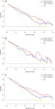
          The area under the receiver‐operator characteristic (ROC) curve (AUC) was used to evaluate the predictive accuracy of the radiomic signature in both the training and test groups. The results of decision curve analysis (DCA) indicated that the radiomic nomogram achieved the most net benefit.

          Results

          The AUC values of ER evaluation for the radiomics signature were 0.80 (training cohort), 0.81 (internal validation cohort), and 0.78 (external validation cohort). Multivariate logistic analysis identified the radiomic signature, CA19‐9 level, and clinical stage as independent parameters of ER. A radiomic nomogram was then developed incorporating the CA19‐9 level and clinical stage. The AUC values for ER risk evaluation using the radiomic nomogram were 0.87 (training cohort), 0.88 (internal validation cohort), and 0.85 (external validation cohort).

          Data Conclusion

          The radiomic nomogram can effectively evaluate ER risks in patients with resectable pancreatic cancer preoperatively, which could potentially improve treatment strategies and facilitate personalized therapy in pancreatic cancer.

          Level of Evidence: 4

          Technical Efficacy: Stage 4

          J. Magn. Reson. Imaging 2020;52:231–245.

          Related collections

          Most cited references30

          • Record: found
          • Abstract: found
          • Article: not found

          Development and Validation of a Radiomics Nomogram for Preoperative Prediction of Lymph Node Metastasis in Colorectal Cancer.

          To develop and validate a radiomics nomogram for preoperative prediction of lymph node (LN) metastasis in patients with colorectal cancer (CRC).
            Bookmark
            • Record: found
            • Abstract: not found
            • Article: not found

            Radiomics Analysis for Evaluation of Pathological Complete Response to Neoadjuvant Chemoradiotherapy in Locally Advanced Rectal Cancer

              Bookmark
              • Record: found
              • Abstract: found
              • Article: not found

              Tumor markers in pancreatic cancer: a European Group on Tumor Markers (EGTM) status report.

              Pancreatic ductal adenocarcinoma is one of the most difficult malignancies to diagnose and treat. The aim of this article is to review how tumor markers can aid the diagnosis and management of patients with this malignancy. The most widely used and best validated marker for pancreatic cancer is CA 19-9. Inadequate sensitivity and specificity limit the use of CA 19-9 in the early diagnosis of pancreatic cancer. In non-jaundiced patients, however, CA 19-9 may complement other diagnostic procedures. In patients with resectable pancreatic cancer, presurgical and postresection CA 19-9 levels correlate with overall survival. In advanced disease, elevated pretreatment levels of CA 19-9 are associated with adverse patient outcome and thus may be combined with other factors for risk stratification. Most, but not all, reports indicate that serial levels of CA 19-9 correlate with response to systemic therapy. Use of CA 19-9 kinetics in conjunction with imaging is therefore recommended in monitoring therapy. Although several potential serum and tissue markers for pancreatic cancer are currently undergoing evaluation, none are sufficiently validated for routine clinical use. CA 19-9 thus remains the serum pancreatic cancer marker against which new markers for this malignancy should be judged.
                Bookmark

                Author and article information

                Contributors
                shirleybai@zju.edu.cn
                liangtingbo@zju.edu.cn
                Journal
                J Magn Reson Imaging
                J Magn Reson Imaging
                10.1002/(ISSN)1522-2586
                JMRI
                Journal of Magnetic Resonance Imaging
                John Wiley & Sons, Inc. (Hoboken, USA )
                1053-1807
                1522-2586
                23 December 2019
                July 2020
                : 52
                : 1 ( doiID: 10.1002/jmri.v52.1 )
                : 231-245
                Affiliations
                [ 1 ] Department of Hepatobiliary and Pancreatic Surgery First Affiliated Hospital, Zhejiang University School of Medicine Hangzhou China
                [ 2 ] Zhejiang Provincial Key Laboratory of Pancreatic Disease Hangzhou China
                [ 3 ] Innovation Center for the Study of Pancreatic Diseases Zhejiang Province China
                [ 4 ] Department of Radiology First Affiliated Hospital, Zhejiang University School of Medicine Hangzhou China
                [ 5 ] Department of Pathology, First Affiliated Hospital Zhejiang University School of Medicine Hangzhou China
                [ 6 ] GE Healthcare China Shanghai China
                [ 7 ] Department of Radiology, Second Affiliated Hospital Zhejiang University School of Medicine Hangzhou China
                Author notes
                [*] [* ]Address reprint requests to: T.‐B.L. or X.‐L.B., 79 Qingchun Road, Hangzhou 310009, China. E‐mail: liangtingbo@ 123456zju.edu.cn or shirleybai@ 123456zju.edu.cn
                [†]

                These authors contributed equally to this work.

                Author information
                https://orcid.org/0000-0003-4825-2189
                https://orcid.org/0000-0002-5942-7282
                Article
                JMRI27024
                10.1002/jmri.27024
                7317738
                31867839
                1cd079cf-b0ed-4a60-a8c6-565df95117fb
                © 2019 The Authors. Journal of Magnetic Resonance Imaging published by Wiley Periodicals, Inc. on behalf of International Society for Magnetic Resonance in Medicine.

                This is an open access article under the terms of the http://creativecommons.org/licenses/by-nc/4.0/ License, which permits use, distribution and reproduction in any medium, provided the original work is properly cited and is not used for commercial purposes.

                History
                : 25 September 2019
                : 27 November 2019
                : 27 November 2019
                Page count
                Figures: 9, Tables: 4, Pages: 15, Words: 7047
                Categories
                Editorial
                Editorials
                Custom metadata
                2.0
                July 2020
                Converter:WILEY_ML3GV2_TO_JATSPMC version:5.8.4 mode:remove_FC converted:26.06.2020

                Radiology & Imaging
                pancreatic cancer,recurrence,relapse,risk assessment,radiomics
                Radiology & Imaging
                pancreatic cancer, recurrence, relapse, risk assessment, radiomics

                Comments

                Comment on this article