- Record: found
- Abstract: found
- Article: found
A Case of Pretibial Epidermolysis Bullosa with Novel Mutations of the COL7A1 Gene
brief-report
Read this article at
There is no author summary for this article yet. Authors can add summaries to their articles on ScienceOpen to make them more accessible to a non-specialist audience.
Abstract
Dear Editor:
Pretibial epidermolysis bullosa (PEB) was first described by Kuske in 19461. The author
reported two cases of PEB with recurrent blisters in a middle-aged man and his son.
PEB is a rare subtype of dominant dystrophic epidermolysis bullosa (DDEB). Symptoms
of DDEB usually appear in infancy, and severe blistering can be life-threatening.
On the other hand, PEB is characterized by mild blistering, erosions, and milia localized
to the shins. The age of onset is variable, and some patients do not develop signs
and symptoms until adulthood. In previous studies, nine patients with mutations in
the COL7A1 gene have been reported2
3
4
5. Herein, we report a case of late-onset PEB and new mutations in the COL7A1 gene.
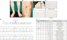
An 86-year-old Japanese male presented with a 1-month history of recurrent blisters
on the shins. Several erythema, tense blisters, erosions, and scars after healing
were present on his shins (Fig. 1A, B). All of his toenails were dystrophic (Fig.
1C). His fingernails, teeth, and hair were not involved. Toenail dystrophy had persisted
from his childhood, and his father also had the condition.
Blood investigations, including a complete blood picture, liver and renal functions,
antinuclear antibody, anti-BP180 antibody, and immunoglobulin (Ig) patterns, reported
normal results.
A skin biopsy showed a subepidermal bulla with poor inflammatory cell infiltration.
The roof of the blister was intact. Mild infiltration of lymphocytes, neutrophils,
and histiocytes was observed (Fig. 1D). At the epidermal side and the dermal side
of the blister, direct immunofluorescence showed no deposition of IgG, IgM, IgA, and
C3. Immunohistochemistry using an anti-collagen type VII monoclonal antibody revealed
that staining was less intense at the basement membrane (Fig. 1E, F). Total RNA was
extracted from peripheral blood, and cDNA was synthesized. Direct sequencing was performed
to detect mutations in the COL7A1 gene. We identified two novel glycine substitution
mutations, namely c.5264G>T (p.Gly1755Val) and c.5345G>C (p.Gly1782Ala), in exons
59 and exon 61, respectively. Both mutations have not been previously reported (Fig.
1G).
After starting treatment with topical corticosteroid and vitamin D3 ointments, no
blisters appeared. Milia occurred after lesions improved with topical benzoyl peroxide.
In the nine cases previously reported, the age of onset of blisters and erosions on
the shins ranged from 1 month to 52 years. Here, we present the oldest age of onset
of blisters and erosions on the shins. Seven of the nine previously reported cases
involved toenail dystrophy (Fig. 1H)2
3
4
5. This case also involved toenail dystrophy that started in childhood. Toenail dystrophy
beginning in childhood may be a clue for PEB diagnosis. We report of two novel glycine
substitution mutations in COL7A1 gene, c.5264G>T (p.Gly1755Val) and c.5345G>C (p.Gly1782Ala)
in exons 59 and 61, respectively, that occurred in late-onset PEB. It is possible
that the both mutations are on the same father-derived allele, but have relatively
low impact on the anchoring fibril formation to generate mild phenotype. Another possibility
is that one mutation is the father-derived dominant mutation and another mutation
might be a silent glycine substitution mutation from his mother.
Related collections
Most cited references5
- Record: found
- Abstract: found
- Article: not found
Pretibial dystrophic epidermolysis bullosa: a recessively inherited COL7A1 splice site mutation affecting procollagen VII processing.
C M Betts, P Posteraro, A. Costa … (1999)
- Record: found
- Abstract: not found
- Article: not found
Splicing mutation in the COL7A1 gene mRNA exon 71 in a female patient with pretibial epidermolysis bullosa.
Tomoko Kitazawa, Tamihiro Kawakami, Maya Matsuoka … (2014)
- Record: found
- Abstract: not found
- Article: not found
Novel missense mutation in a patient with recessive pretibial epidermolysis bullosa and a mild phenotype
S. Menzies, L. Liu, L. Cunningham … (2015)