6
views
0
recommends
+1 Recommend
0 collections
    0
    shares
      • Record: found
      • Abstract: found
      • Article: found
      Is Open Access

      Expression of GLP-1 receptor and CD26 in human thyroid C-cells: The association of thyroid C-cell tumorigenesis with incretin-based medicine

      research-article

      Read this article at

      Bookmark
          There is no author summary for this article yet. Authors can add summaries to their articles on ScienceOpen to make them more accessible to a non-specialist audience.

          Abstract

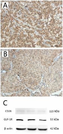
          Recent reports have demonstrated that long-term and high dosage treatments with incretin-based medicine, such as hormone glucagon-like peptide-1 (GLP-1) may induce thyroid C-cell pathological changes in rodents, rather than in humans. Doubts regarding the tumorigenic potential of GLP-1 analogues in human thyroid C-cells remain. The present study aimed to determine the expression levels of GLP-1 receptor (GLP-1R) and cluster of differentiation 26 (CD26) in the C-cells of thyroid tissues from non-neoplastic, medullary carcinoma and hyperplasia subjects, and to explore the potential clinical significance. The following cases were analyzed: Medullary thyroid carcinoma (n=62, including 59 paraffin-embedded samples and 3 fresh frozen samples), C-cell hyperplasia (n=20, paraffin-embedded samples) and non-neoplastic thyroid tissue samples (n=7, paraffin-embedded samples). GLP-1R and CD26 expression was detected using immunohistochemical staining and western blotting. There were significant differences in the expression levels of the two markers between medullary thyroid carcinoma and C-cell hyperplasia, in addition to between medullary thyroid carcinoma and non-neoplastic thyroid tissue following immunohistochemical staining. Similar significant differences in the expression of GLP-1R and CD26 were detected using western blot analysis in the medullary thyroid carcinoma compared with non-neoplastic thyroid tissue sectioned from the aforementioned fresh frozen samples. There was a significant negative correlation between GLP-1R and CD26 expression. In addition, the present data indicated that GLP-1R expression was associated with the age of the patients with medullary thyroid carcinoma. These results suggested that GLP-1R and CD26 may be closely associated with the development of thyroid C-cell hyperplasia and medullary thyroid carcinoma, and indicated the importance of being aware of the side effects of incretin medicine.

          Related collections

          Most cited references28

          • Record: found
          • Abstract: found
          • Article: not found

          Dipeptidyl-peptidase IV (CD26)--role in the inactivation of regulatory peptides.

          Dipeptidyl-peptidase IV (DPP IV/CD26) has a dual function as a regulatory protease and as a binding protein. Its role in the inactivation of bioactive peptides was recognized 20 years ago due to its unique ability to liberate Xaa-Pro or Xaa-Ala dipeptides from the N-terminus of regulatory peptides, but further examples are now emerging from in vitro and vivo experiments. Despite the minimal N-terminal truncation by DPP IV, many mammalian regulatory peptides are inactivated--either totally or only differentially--for certain receptor subtypes. Important DPP IV substrates include neuropeptides like neuropeptide Y or endomorphin, circulating peptide hormones like peptide YY, growth hormone-releasing hormone, glucagon-like peptides(GLP)-1 and -2, gastric inhibitory polypeptide as well as paracrine chemokines like RANTES (regulated on activation normal T cell expressed and secreted), stromal cell-derived factor, eotaxin and macrophage-derived chemokine. Based on these findings the potential clinical uses of selective DPP IV inhibitors or DPP IV-resistant analogues, especially for the insulinotropic hormone GLP-1, have been tested to enhance insulin secretion and to improve glucose tolerance in diabetic animals. Thus, DPP IV appears to be a major physiological regulator for some regulatory peptides, neuropeptides, circulating hormones and chemokines.
            Bookmark
            • Record: found
            • Abstract: found
            • Article: not found

            Glucagon-like Peptide-1 receptor agonists activate rodent thyroid C-cells causing calcitonin release and C-cell proliferation.

            Liraglutide is a glucagon-like peptide-1 (GLP-1) analog developed for type 2 diabetes. Long-term liraglutide exposure in rodents was associated with thyroid C-cell hyperplasia and tumors. Here, we report data supporting a GLP-1 receptor-mediated mechanism for these changes in rodents. The GLP-1 receptor was localized to rodent C-cells. GLP-1 receptor agonists stimulated calcitonin release, up-regulation of calcitonin gene expression, and subsequently C-cell hyperplasia in rats and, to a lesser extent, in mice. In contrast, humans and/or cynomolgus monkeys had low GLP-1 receptor expression in thyroid C-cells, and GLP-1 receptor agonists did not activate adenylate cyclase or generate calcitonin release in primates. Moreover, 20 months of liraglutide treatment (at >60 times human exposure levels) did not lead to C-cell hyperplasia in monkeys. Mean calcitonin levels in patients exposed to liraglutide for 2 yr remained at the lower end of the normal range, and there was no difference in the proportion of patients with calcitonin levels increasing above the clinically relevant cutoff level of 20 pg/ml. Our findings delineate important species-specific differences in GLP-1 receptor expression and action in the thyroid. Nevertheless, the long-term consequences of sustained GLP-1 receptor activation in the human thyroid remain unknown and merit further investigation.
              Bookmark
              • Record: found
              • Abstract: found
              • Article: not found

              Tissue-specific expression of the human receptor for glucagon-like peptide-I: brain, heart and pancreatic forms have the same deduced amino acid sequences.

              Y Wei, S Mojsov (1995)
              Glucagon-like peptide-I(GLP-I), encoded by the glucagon gene and released from the gut in response to nutrients, is a potent stimulator of glucose-induced insulin secretion. In human subjects GLP-I exerts its physiological effect as an incretin. The incretin effect of GLP-I is preserved in patients with Type II diabetes mellitus (NIDDM), suggesting that GLP-I receptor agonist can be used therapeutically in this group of patients. In these studies we addressed the question of whether GLP-I has broader actions in human physiology. To investigate this issue we examined the tissue distribution of GLP-I receptor using RNAse protection assay in order to avoid the cross-reactivities with structurally related receptors and to increase the sensitivity of detection. The riboprobe was synthesized from the human pancreatic GLP-I receptor cDNA and used in hybridization experiments with total RNA isolated from different human tissues. In addition to the pancreas, we found expression of GLP-I receptor mRNA in lung, brain, kidney, stomach and heart. Peripheral tissues which are the major sites of glucose turnover, such as liver, skeletal muscle and adipose did not express the pancreatic form of the GLP-I receptor. We also cloned and sequenced GLP-I receptor cDNA from human brain and heart. The deduced amino acid sequences are the same as the sequence found in the pancreas. These results indicate that GLP-I might have effects beyond the pancreas, including the cardiovascular and central nervous systems where a receptor with the same ligand binding specificity is found.
                Bookmark

                Author and article information

                Journal
                Oncol Lett
                Oncol Lett
                OL
                Oncology Letters
                D.A. Spandidos
                1792-1074
                1792-1082
                April 2017
                20 February 2017
                20 February 2017
                : 13
                : 4
                : 2684-2690
                Affiliations
                [1 ]Department of Endocrinology, The First Hospital of Harbin Medical University, Harbin, Heilongjiang 150001, P.R. China
                [2 ]Department of Pathology, The First Hospital of Harbin Medical University, Harbin, Heilongjiang 150001, P.R. China
                [3 ]Department of Pathology, The Third Hospital of Harbin Medical University, Harbin, Heilongjiang 150001, P.R. China
                Author notes
                Correspondence to: Professor Xiaomin Liu, Department of Endocrinology, The First Hospital of Harbin Medical University, 23 Youzheng Street, Nangang, Harbin, Heilongjiang 150001, P.R. China, E-mail: 1344230@ 123456qq.com
                Article
                OL-0-0-5752
                10.3892/ol.2017.5752
                5403711
                1d3a4ff2-c7bc-461a-ab5e-d6db9f61757d
                Copyright: © Song et al.

                This is an open access article distributed under the terms of the Creative Commons Attribution-NonCommercial-NoDerivs License, which permits use and distribution in any medium, provided the original work is properly cited, the use is non-commercial and no modifications or adaptations are made.

                History
                : 08 August 2015
                : 20 December 2016
                Categories
                Articles

                Oncology & Radiotherapy
                medullary thyroid cancer,glucagon-like peptide-1 receptor,cluster of differentiation 26,thyroid c-cell

                Comments

                Comment on this article