2
views
0
recommends
+1 Recommend
0 collections
    0
    shares
      • Record: found
      • Abstract: found
      • Article: found
      Is Open Access

      Assessment of Solid Pulmonary Nodules or Masses Using Zero Echo Time MR Lung Imaging: A Prospective Head-to-Head Comparison With CT

      research-article

      Read this article at

      Bookmark
          There is no author summary for this article yet. Authors can add summaries to their articles on ScienceOpen to make them more accessible to a non-specialist audience.

          Abstract

          Objective

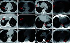
          The aim of this study is to determine the potential of zero echo time (ZTE) MR lung imaging in the assessment of solid pulmonary nodules or masses and diagnostic consistency to CT in terms of morphologic characterization.

          Methods

          Our Institutional Review Board approved this prospective study. Seventy-one patients with solid pulmonary nodules or masses larger than 1 cm in diameter confirmed by chest CT were enrolled and underwent further lung ZTE-MRI scans within 7 days. ZTE-MRI and CT images were compared in terms of image quality and imaging features. Unidimensional diameter and three-dimensional volume measurements on both modalities were manually measured and compared using the Wilcoxon signed-rank test, intraclass correlation coefficient (ICC), Pearson’s correlation analysis, and Bland–Altman analysis. Multivariable logistic regression analysis was used to identify the factors associated with significant inter-modality variation of volume.

          Results

          Fifty-four of 71 (76.1%) patients were diagnosed with lung cancer. Subjective image quality was superior in CT compared with ZTE-MRI ( p < 0.001). Inter-modality agreement for the imaging features was moderate for emphysema (kappa = 0.50), substantial for fibrosis (kappa = 0.76), and almost perfect (kappa = 0.88-1.00) for the remaining features. The size measurements including diameter and volume between ZTE-MRI and CT showed no significant difference ( p = 0.36 for diameter and 0.60 for volume) and revealed perfect inter-observer (ICC = 0.975–0.980) and inter-modality (ICC = 0.942–0.992) agreements. Multivariable analysis showed that non-smooth margin [odds ratio (OR) = 6.008, p = 0.015] was an independent predictor for the significant inter-modality variation of volume.

          Conclusion

          ZTE lung imaging is feasible as a part of chest MRI in the assessment and surveillance for solid pulmonary nodules or masses larger than 1 cm, presenting perfect agreement with CT in terms of morphologic characterization.

          Related collections

          Most cited references31

          • Record: found
          • Abstract: found
          • Article: not found

          User-guided 3D active contour segmentation of anatomical structures: significantly improved efficiency and reliability.

          Active contour segmentation and its robust implementation using level set methods are well-established theoretical approaches that have been studied thoroughly in the image analysis literature. Despite the existence of these powerful segmentation methods, the needs of clinical research continue to be fulfilled, to a large extent, using slice-by-slice manual tracing. To bridge the gap between methodological advances and clinical routine, we developed an open source application called ITK-SNAP, which is intended to make level set segmentation easily accessible to a wide range of users, including those with little or no mathematical expertise. This paper describes the methods and software engineering philosophy behind this new tool and provides the results of validation experiments performed in the context of an ongoing child autism neuroimaging study. The validation establishes SNAP intrarater and interrater reliability and overlap error statistics for the caudate nucleus and finds that SNAP is a highly reliable and efficient alternative to manual tracing. Analogous results for lateral ventricle segmentation are provided.
            Bookmark
            • Record: found
            • Abstract: found
            • Article: not found

            RECIST 1.1-Update and clarification: From the RECIST committee.

            The Response Evaluation Criteria in Solid Tumours (RECIST) were developed and published in 2000, based on the original World Health Organisation guidelines first published in 1981. In 2009, revisions were made (RECIST 1.1) incorporating major changes, including a reduction in the number of lesions to be assessed, a new measurement method to classify lymph nodes as pathologic or normal, the clarification of the requirement to confirm a complete response or partial response and new methodologies for more appropriate measurement of disease progression. The purpose of this paper was to summarise the questions posed and the clarifications provided as an update to the 2009 publication.
              Bookmark
              • Record: found
              • Abstract: found
              • Article: not found

              CT Radiogenomic Characterization of EGFR, K-RAS, and ALK Mutations in Non-Small Cell Lung Cancer.

              To assess the association between CT features and EGFR, ALK, KRAS mutations in non-small cell lung cancer.
                Bookmark

                Author and article information

                Contributors
                Journal
                Front Oncol
                Front Oncol
                Front. Oncol.
                Frontiers in Oncology
                Frontiers Media S.A.
                2234-943X
                26 April 2022
                2022
                : 12
                : 812014
                Affiliations
                [1] 1 Department of Medical Imaging, Yueyang Central Hospital , Yueyang, China
                [2] 2 Department of Radiology, The Third Xiangya Hospital, Central South University , Changsha, China
                [3] 3 Magnetic Resonance (MR) Research, General Electric (GE) Healthcare , Beijing, China
                Author notes

                Edited by: Takahito Nakajima, University of Tsukuba, Japan

                Reviewed by: Sikandar Shaikh, Shadan Hospital and Institute of Medical Sciences, India; Beigelman Catherine, Centre Hospitalier Universitaire Vaudois (CHUV), Switzerland; Gael Dournes, Centre Hospitalier Universitaire de Bordeaux, France

                *Correspondence: Wenming Zhou, xy31929@ 123456163.com

                †These authors have contributed equally to this work

                This article was submitted to Cancer Imaging and Image-directed Interventions, a section of the journal Frontiers in Oncology

                Article
                10.3389/fonc.2022.812014
                9088008
                35558517
                1f5ce929-3389-45d0-8089-2fe11b502bc6
                Copyright © 2022 Liu, Feng, Liu, Fu, He, Cheng, Mao and Zhou

                This is an open-access article distributed under the terms of the Creative Commons Attribution License (CC BY). The use, distribution or reproduction in other forums is permitted, provided the original author(s) and the copyright owner(s) are credited and that the original publication in this journal is cited, in accordance with accepted academic practice. No use, distribution or reproduction is permitted which does not comply with these terms.

                History
                : 17 November 2021
                : 24 March 2022
                Page count
                Figures: 7, Tables: 5, Equations: 0, References: 31, Pages: 11, Words: 5015
                Categories
                Oncology
                Original Research

                Oncology & Radiotherapy
                magnetic resonance imaging,zero echo time sequence,pulmonary nodule,comparison,computed tomography

                Comments

                Comment on this article