24
views
0
recommends
+1 Recommend
0 collections
    0
    shares
      • Record: found
      • Abstract: found
      • Article: found
      Is Open Access

      Balloon-occluded retrograde transvenous obliteration for gastric varices: the relationship between the clinical outcome and gastrorenal shunt occlusion

      research-article

      Read this article at

      Bookmark
          There is no author summary for this article yet. Authors can add summaries to their articles on ScienceOpen to make them more accessible to a non-specialist audience.

          Abstract

          Background

          The rupture of gastric varices is associated with high mortality rate. Balloon-occluded retrograde transvenous obliteration (B-RTO), a minimally invasive procedure that was introduced in the mid-1990s, has been widely accepted in Japan. Several reports have indicated that B-RTO yields satisfactory results; however, few reports have discussed the recurrence of gastric varices after this therapy. The purpose of this study is to retrospectively evaluate the technical aspects of B-RTO and the recurrence of gastric varices after treatment with this procedure.

          Methods

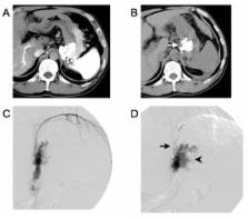
          B-RTO was performed in 47 patients with gastric varices, who were at a risk of variceal ruptures and who may or may not have had a history of variceal bleeding. We injected a sclerosing agent into the gastric varices for 30-60 minutes. To evaluate the therapeutic efficacy of the technique, we obtained contrast-enhanced computed tomography (CT) scans 5 days after B-RTO. As a general rule, if the gastric varices did not appear thrombosed, we repeated the procedure 7 days after the first procedure.

          Results

          B-RTO was a technical success in 37 patients. It was performed once in 26 patients, twice in 6 patients, thrice in 2 patients, and 4 times in 3 patients. Contrast-enhanced CT scans obtained after B-RTO showed thrombosed gastrorenal shunts in 29 patients and patent gastrorenal shunts in 8 patients. The gastric varices recurred in 2 patients who had patent gastrorenal shunts. The overall cumulative relapse-free rate of gastric varices was 90% at 5 years after B-RTO.

          Conclusions

          B-RTO is an effective treatment modality for gastric varices. Moreover, obliteration of the gastrorenal shunt as well as the gastric varices appears to be important for the treatment of gastric varices.

          Related collections

          Most cited references12

          • Record: found
          • Abstract: found
          • Article: not found

          Prevalence, classification and natural history of gastric varices: a long-term follow-up study in 568 portal hypertension patients.

          To determine the prevalence and natural history of gastric varices, we prospectively studied 568 patients (393 bleeders and 175 nonbleeders) with portal hypertension (cirrhosis in 301 patients, noncirrhotic portal fibrosis in 115 patients, extrahepatic portal vein obstruction in 117 patients and hepatic venous outflow obstruction in 35 patients). Primary (present at initial examination) gastric varices were seen in 114 (20%) patients; more were present in bleeders than in non-bleeders (27% vs. 4%, respectively; p < 0.001). Secondary (occurring after obliteration of esophageal varices) gastric varices developed in 33 (9%) patients during follow-up of 24.6 +/- 5.3 mo. Gastric varices (compared with esophageal varices) bled in significantly fewer patients (25% vs. 64%, respectively). Gastric varices had a lower bleeding risk factor than did esophageal varices (2.0 +/- 0.5 vs. 4.3 +/- 0.4, respectively) but bled more severely (4.8 +/- 0.6 vs. 2.9 +/- 0.3 transfusion units per patient, respectively). Once a varix bled, mortality was more likely (45%) in gastric varix patients. Gastric varices were classified as gastroesophageal or isolated gastric varices. Type 1 gastroesophageal varices (lesser curve varices) were the most common (75%). After obliteration of esophageal varices, type 1 gastroesophageal varices disappeared in 59% of patients and persisted in the remainder; bleeding from persistent gastroesophageal varices was more common than it was from gastroesophageal varices that were obliterated (28% vs. 2%, respectively; p < 0.001). Type 2 gastroesophageal varices, which extend to greater curvature, bled often (55%) and were associated with high mortality. Type 1 isolated gastric varices patients had only fundal varices, with a high (78%) incidence of bleeding.(ABSTRACT TRUNCATED AT 250 WORDS)
            Bookmark
            • Record: found
            • Abstract: found
            • Article: not found

            Retrograde transvenous obliteration of gastric varices.

            To evaluate the clinical efficacy, techniques, and complications associated with balloon-occluded retrograde transvenous obliteration of gastric varices. Between December 1994 and November 1997, balloon-occluded retrograde transvenous obliteration was performed on 20 patients with gastric varices in danger of rupture and with gastrorenal shunts; three patients also had hepatic encephalopathy. The sclerosant was injected into the gastric varices during balloon occlusion. The degree of progression of the gastric varices and of collateral veins was classified into five grades, with grade 1 being least progression and grade 5 most progression; collateral veins that had developed were treated with embolization. Follow-up consisted of fiberoptic endoscopy and computed tomography. Technical success was achieved in all patients. Occlusion of collateral veins was essential for the occlusion of gastric varices with a grade greater than grade 2. The clinical symptoms of hepatic encephalopathy in the three patients improved remarkably. Follow-up endoscopy 3 months after the procedure revealed the disappearance of gastric varices in 15 patients and reduced variceal size in five. During the follow-up period, 19 patients had no recurrence of gastric varices; three patients had aggravation of the esophageal varices. Balloon-occluded retrograde transvenous obliteration is a feasible alternative to a transjugular intrahepatic portosystemic shunt for patients with large gastrorenal shunts or hepatic encephalopathy (or both).
              Bookmark
              • Record: found
              • Abstract: found
              • Article: not found

              Endoscopic injection sclerosis in bleeding gastric varices.

              Ninety-two consecutive, nonrandomized patients with bleeding varices were prospectively studied using sclerotherapy to control and prevent rebleeding. During this study, nine patients with gastric variceal bleeding were identified. A gastric variceal subset is defined and represents a 10% incidence in this series. All patients presented with indexed gastric bleeding varices that subsequently accounted for 34 bleeding sessions. The units of blood per rebleeding episode, hospital days, cost, and outcome were markedly different from the esophageal variceal groups. Initial management of indexed bleeding episodes by sclerotherapy and Sengstaken-Blakemore tubes were comparable; however, the number of rebleeding episodes was much higher. There was poor control of rebleeding with an associated higher rebleeding mortality and complications secondary to repeated sclerotherapy and Sengstaken-Blakemore tube use. In 37% of the patients, rebleeding was the direct result of gastric ulcerations at the endoscopic injection sclerotherapy site. The survival curve of this group was much lower than esophageal variceal bleeders. Endoscopic injection sclerotherapy in patients with bleeding gastric varices offers only temporary control of bleeding, and the high incidence of severe early rebleeding requires consideration of alternative methods for management or modified sclerotherapy techniques.
                Bookmark

                Author and article information

                Journal
                BMC Med Imaging
                BMC Medical Imaging
                BioMed Central
                1471-2342
                2010
                14 January 2010
                : 10
                : 2
                Affiliations
                [1 ]Department of Radiology, Iwate Medical University, Morioka, Japan
                [2 ]Department of Radiology, Morioka Red Cross Hospital, Morioka, Japan
                [3 ]Department of Critical Care Medicine, Iwate Medical University, Morioka, Japan
                Article
                1471-2342-10-2
                10.1186/1471-2342-10-2
                2820437
                20074342
                241e41d5-472c-4b14-8f53-a527937e1aae
                Copyright ©2010 Katoh et al; licensee BioMed Central Ltd.

                This is an Open Access article distributed under the terms of the Creative Commons Attribution License ( http://creativecommons.org/licenses/by/2.0), which permits unrestricted use, distribution, and reproduction in any medium, provided the original work is properly cited.

                History
                : 16 January 2009
                : 14 January 2010
                Categories
                Research article

                Radiology & Imaging
                Radiology & Imaging

                Comments

                Comment on this article