7
views
0
recommends
+1 Recommend
0 collections
    0
    shares
      • Record: found
      • Abstract: found
      • Article: found
      Is Open Access

      Role of autophagy in tumor necrosis factor-α-induced apoptosis of osteoblast cells

      research-article

      Read this article at

      Bookmark
          There is no author summary for this article yet. Authors can add summaries to their articles on ScienceOpen to make them more accessible to a non-specialist audience.

          Abstract

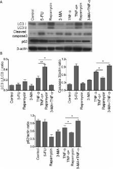
          The aim of this study is to investigate the role of tumor necrosis factor-α (TNF-α) in apoptosis and autophagy of mouse osteoblast MC3T3-E1 cells, as well as the crosstalk between autophagy and apoptosis. Mouse osteoblast MC3T3-E1 cells were cultured in vitro and treated with 5-fluorouracil (5-FU), rapamycin, 3-methyl adenine (3-MA) and TNF-α either alone or in combination, respectively. MTT assays were used to monitor the cell viability upon different treatments. Annexin-V-FITC/propidium iodide (PI) staining was used to detect the apoptotic rate of osteoblasts. Autophagic structure and apoptotic bodies were visualized by transmission electron microscopy (TEM). Western blot analysis was performed to detect the autophagic marker LC3-II/I, p62 and apoptotic marker cleaved caspase-3. TNF-α inhibits MC3T3-E1 cell viability in a dose-dependent and time-dependent manner. Annexin-V-FITC/PI staining, coupled with TEM, showed that TNF-α induced cell apoptosis and autophagy in MC3T3-E1 cells. The autophagy inducer rapamycin ameliorated TNF-α-induced apoptosis. In contrast, 3-MA, which is an autophagy inhibitor, caused an exaggerated induction of TNF-α-induced apoptosis. TNF-α upregulated autophagy marker LC3-II/I, but downregulated p62 in osteoblasts. Combined treatment of rapamycin and TNF-α further exaggerated this effect, whereas co-treatment of 3-MA and TNF-α decreased LC3-II/I, but increased p62 compared with TNF-α alone. In addition, TNF-α caused an induction of apoptotic marker cleaved caspase-3. TNF-α-mediated induction of cleaved caspase-3 was downregulated by rapamycin, but upregulated by 3-MA, respectively. TNF-α induced both autophagy and apoptosis in osteoblasts, and upregulated autophagy protects the cell by reducing TNF-α-induced apoptosis.

          Related collections

          Most cited references16

          • Record: found
          • Abstract: found
          • Article: not found

          Autophagy regulates TNFα-mediated joint destruction in experimental arthritis.

          Autophagy is a homeostatic process to recycle dispensable and damaged cell organelles. Dysregulation of autophagic pathways has recently been implicated in the pathogenesis of various diseases. Here, we investigated the role of autophagy during joint destruction in arthritis. Autophagy in osteoclasts was analysed in vitro and ex vivo by transmission electron microscopy, Western blotting and immunohistochemistry for Beclin1 and Atg7. Small molecule inhibitors, LysMCre-mediated knockout of Atg7 and lentiviral overexpression of Beclin1 were used to modulate autophagy in vitro and in vivo. Osteoclast differentiation markers were quantified by real-time PCR. The extent of bone and cartilage destruction was analysed in human tumour necrosis factor α transgenic (hTNFα tg) mice after adoptive transfer with myeloid specific Atg7-deficient bone marrow. Autophagy was activated in osteoclasts of human rheumatoid arthritis (RA) showing increased expression of Beclin1 and Atg7. TNFα potently induced the expression of autophagy-related genes and activated autophagy in vitro and in vivo. Activation of autophagy by overexpression of Beclin1-induced osteoclastogenesis and enhanced the resorptive capacity of cultured osteoclasts, whereas pharmacologic or genetic inactivation of autophagy prevented osteoclast differentiation. Arthritic hTNFα tg mice transplanted with Atg7(fl/fl)×LysMCre(+) bone marrow cells (BMC) showed reduced numbers of osteoclasts and were protected from TNFα-induced bone erosion, proteoglycan loss and chondrocyte death. These findings demonstrate that autophagy is activated in RA in a TNFα-dependent manner and regulates osteoclast differentiation and bone resorption. We thus provide evidence for a central role of autophagy in joint destruction in RA.
            Bookmark
            • Record: found
            • Abstract: found
            • Article: not found

            Osteoblast programmed cell death (apoptosis): modulation by growth factors and cytokines.

            Once osteoblasts have completed their bone-forming function, they are either entrapped in bone matrix and become osteocytes or remain on the surface as lining cells. Nonetheless, 50-70% of the osteoblasts initially present at the remodeling site cannot be accounted for after enumeration of lining cells and osteocytes. We hypothesized that the missing osteoblasts die by apoptosis and that growth factors and cytokines produced in the bone microenvironment influence this process. We report that murine osteoblastic MC3T3-E1 cells underwent apoptosis following removal of serum, or addition of tumor necrosis factor (TNF), as indicated by terminal deoxynucleotidyl transferase-mediated dUTP-nick end labeling and DNA fragmentation studies. Transforming growth factor-beta and interleukin-6 (IL-6)-type cytokines had antiapoptotic effects because they were able to counteract the effect of serum starvation or TNF. In addition, anti-Fas antibody stimulated apoptosis of human osteoblastic MG-63 cells and IL-6-type cytokines prevented these changes. The induction of apoptosis in MG-63 cells was associated with an increase in the ratio of the proapoptotic protein bax to the antiapoptotic protein bcl-2, and oncostatin M prevented this change. Examination of undecalcified sections of murine cancellous bone revealed the presence of apoptotic cells, identified as osteoblasts by their proximity to osteoid seams and their juxtaposition to cuboidal osteoblasts. Assuming an osteoblast life span of 300 h and a prevalence of apoptosis of 0.6%, we calculated that the fraction that undergo this process in vivo can indeed account for the missing osteoblasts. These findings establish that osteoblasts undergo apoptosis and strongly suggest that the process can be modulated by growth factors and cytokines produced in the bone microenvironment.
              Bookmark
              • Record: found
              • Abstract: found
              • Article: not found

              Infection, inflammation, and bone regeneration: a paradoxical relationship.

              Various strategies have been developed to promote bone regeneration in the craniofacial region. Most of these interventions utilize implantable materials or devices. Infections resulting from colonization of these implants may result in local tissue destruction in a manner analogous to periodontitis. This destruction is mediated via the expression of various inflammatory mediators and tissue-destructive enzymes. Given the well-documented association among microbial biofilms, inflammatory mediators, and tissue destruction, it seems reasonable to assume that inflammation may interfere with bone healing and regeneration. Paradoxically, recent evidence also suggests that the presence of certain pro-inflammatory mediators is actually required for bone healing. Bone injury (e.g., subsequent to a fracture or surgical intervention) is followed by a choreographed cascade of events, some of which are dependent upon the presence of pro-inflammatory mediators. If inflammation resolves promptly, then proper bone healing may occur. However, if inflammation persists (which might occur in the presence of an infected implant or graft material), then the continued inflammatory response may result in suboptimal bone formation. Thus, the effect of a given mediator is dependent upon the temporal context in which it is expressed. Better understanding of this temporal sequence may be used to optimize regenerative outcomes.
                Bookmark

                Author and article information

                Journal
                J Investig Med
                J. Investig. Med
                jim
                jim
                Journal of Investigative Medicine
                Journal of Investigative Medicine (BMA House, Tavistock Square, London, WC1H 9JR )
                1081-5589
                1708-8267
                August 2017
                20 June 2017
                : 65
                : 6
                : 1014-1020
                Affiliations
                [1] departmentDepartment of Orthopeadics , The Second Xiangya Hospital, Central South University , Changsha, Hunan, China
                Author notes
                [Correspondence to ] Dr Liwen Zheng, Department of Orthopeadics, The Second Xiangya Hospital, Central South University, Changsha, Hunan 410011, China; zheng_liwen@ 123456yahoo.com
                Author information
                http://orcid.org/0000-0002-7018-9502
                Article
                jim-2017-000426
                10.1136/jim-2017-000426
                5537511
                28634253
                2678d36b-bf30-4d84-b934-0c181947c137
                © American Federation for Medical Research (unless otherwise stated in the text of the article) 2017. All rights reserved. No commercial use is permitted unless otherwise expressly granted.

                This is an Open Access article distributed in accordance with the Creative Commons Attribution Non Commercial (CC BY-NC 4.0) license, which permits others to distribute, remix, adapt, build upon this work non-commercially, and license their derivative works on different terms, provided the original work is properly cited and the use is non-commercial. See: http://creativecommons.org/licenses/by-nc/4.0/

                History
                : 29 January 2017
                : 07 April 2017
                : 13 April 2017
                Categories
                Basic Science/Genetics/Molecular Medicine
                1506
                Original research
                Custom metadata
                unlocked

                tumor necrosis factor-alpha,osteoblasts,antibiotics,apoptosis

                Comments

                Comment on this article