23
views
0
recommends
+1 Recommend
0 collections
    0
    shares
      • Record: found
      • Abstract: found
      • Article: found
      Is Open Access

      ImageJS: Personalized, participated, pervasive, and reproducible image bioinformatics in the web browser

      research-article

      Read this article at

      Bookmark
          There is no author summary for this article yet. Authors can add summaries to their articles on ScienceOpen to make them more accessible to a non-specialist audience.

          Abstract

          Background:

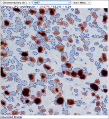
          Image bioinformatics infrastructure typically relies on a combination of server-side high-performance computing and client desktop applications tailored for graphic rendering. On the server side, matrix manipulation environments are often used as the back-end where deployment of specialized analytical workflows takes place. However, neither the server-side nor the client-side desktop solution, by themselves or combined, is conducive to the emergence of open, collaborative, computational ecosystems for image analysis that are both self-sustained and user driven.

          Materials and Methods:

          ImageJS was developed as a browser-based webApp, untethered from a server-side backend, by making use of recent advances in the modern web browser such as a very efficient compiler, high-end graphical rendering capabilities, and I/O tailored for code migration.

          Results:

          Multiple versioned code hosting services were used to develop distinct ImageJS modules to illustrate its amenability to collaborative deployment without compromise of reproducibility or provenance. The illustrative examples include modules for image segmentation, feature extraction, and filtering. The deployment of image analysis by code migration is in sharp contrast with the more conventional, heavier, and less safe reliance on data transfer. Accordingly, code and data are loaded into the browser by exactly the same script tag loading mechanism, which offers a number of interesting applications that would be hard to attain with more conventional platforms, such as NIH's popular ImageJ application.

          Conclusions:

          The modern web browser was found to be advantageous for image bioinformatics in both the research and clinical environments. This conclusion reflects advantages in deployment scalability and analysis reproducibility, as well as the critical ability to deliver advanced computational statistical procedures machines where access to sensitive data is controlled, that is, without local “download and installation”.

          Related collections

          Most cited references17

          • Record: found
          • Abstract: found
          • Article: not found

          Reproducible research in computational science.

          Roger Peng (2011)
          Computational science has led to exciting new developments, but the nature of the work has exposed limitations in our ability to evaluate published findings. Reproducibility has the potential to serve as a minimum standard for judging scientific claims when full independent replication of a study is not possible.
            Bookmark
            • Record: found
            • Abstract: found
            • Article: found
            Is Open Access

            ImmunoRatio: a publicly available web application for quantitative image analysis of estrogen receptor (ER), progesterone receptor (PR), and Ki-67

            Introduction Accurate assessment of estrogen receptor (ER), progesterone receptor (PR), and Ki-67 is essential in the histopathologic diagnostics of breast cancer. Commercially available image analysis systems are usually bundled with dedicated analysis hardware and, to our knowledge, no easily installable, free software for immunostained slide scoring has been described. In this study, we describe a free, Internet-based web application for quantitative image analysis of ER, PR, and Ki-67 immunohistochemistry in breast cancer tissue sections. Methods The application, named ImmunoRatio, calculates the percentage of positively stained nuclear area (labeling index) by using a color deconvolution algorithm for separating the staining components (diaminobenzidine and hematoxylin) and adaptive thresholding for nuclear area segmentation. ImmunoRatio was calibrated using cell counts defined visually as the gold standard (training set, n = 50). Validation was done using a separate set of 50 ER, PR, and Ki-67 stained slides (test set, n = 50). In addition, Ki-67 labeling indexes determined by ImmunoRatio were studied for their prognostic value in a retrospective cohort of 123 breast cancer patients. Results The labeling indexes by calibrated ImmunoRatio analyses correlated well with those defined visually in the test set (correlation coefficient r = 0.98). Using the median Ki-67 labeling index (20%) as a cutoff, a hazard ratio of 2.2 was obtained in the survival analysis (n = 123, P = 0.01). ImmunoRatio was shown to adapt to various staining protocols, microscope setups, digital camera models, and image acquisition settings. The application can be used directly with web browsers running on modern operating systems (e.g., Microsoft Windows, Linux distributions, and Mac OS). No software downloads or installations are required. ImmunoRatio is open source software, and the web application is publicly accessible on our website. Conclusions We anticipate that free web applications, such as ImmunoRatio, will make the quantitative image analysis of ER, PR, and Ki-67 easy and straightforward in the diagnostic assessment of breast cancer specimens.
              Bookmark
              • Record: found
              • Abstract: not found
              • Article: not found

              Computer science. Beyond the data deluge.

                Bookmark

                Author and article information

                Journal
                J Pathol Inform
                JPI
                Journal of Pathology Informatics
                Medknow Publications & Media Pvt Ltd (India )
                2153-3539
                2153-3539
                2012
                20 July 2012
                : 3
                : 25
                Affiliations
                [1]Division Informatics, University of Alabama at Birmingham, Alabama, USA
                [1 ]Division Informatics, Department of Pathology, University of Alabama at Birmingham, Alabama, USA
                [2 ]Division Informatics, Department of Biomedical Engineering, University of Alabama at Birmingham, Alabama, USA
                [3 ]Division Informatics, Department of Electrical and Engineering, University of Alabama at Birmingham, Alabama, USA
                Author notes
                [* ]Corresponding author
                Article
                JPI-3-25
                10.4103/2153-3539.98813
                3424663
                22934238
                2770bf58-0738-478e-9e7a-5ad352b4892c
                Copyright: © 2012 Almeida JS.

                This is an open-access article distributed under the terms of the Creative Commons Attribution License, which permits unrestricted use, distribution, and reproduction in any medium, provided the original author and source are credited.

                History
                : 06 April 2012
                : 06 June 2012
                Categories
                Research Article

                Pathology
                image analysis,webapp,cloud computing
                Pathology
                image analysis, webapp, cloud computing

                Comments

                Comment on this article