52
views
0
recommends
+1 Recommend
0 collections
    0
    shares
      • Record: found
      • Abstract: found
      • Article: not found

      Quantification of the Temporal Evolution of Collagen Orientation in Mechanically Conditioned Engineered Cardiovascular Tissues

      research-article

      Read this article at

      ScienceOpenPublisherPMC
      Bookmark
          There is no author summary for this article yet. Authors can add summaries to their articles on ScienceOpen to make them more accessible to a non-specialist audience.

          Abstract

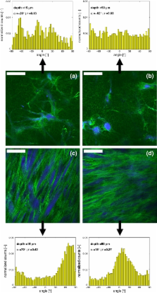
          Load-bearing soft tissues predominantly consist of collagen and exhibit anisotropic, non-linear visco-elastic behavior, coupled to the organization of the collagen fibers. Mimicking native mechanical behavior forms a major goal in cardiovascular tissue engineering. Engineered tissues often lack properly organized collagen and consequently do not meet in vivo mechanical demands. To improve collagen architecture and mechanical properties, mechanical stimulation of the tissue during in vitro tissue growth is crucial. This study describes the evolution of collagen fiber orientation with culture time in engineered tissue constructs in response to mechanical loading. To achieve this, a novel technique for the quantification of collagen fiber orientation is used, based on 3D vital imaging using multiphoton microscopy combined with image analysis. The engineered tissue constructs consisted of cell-seeded biodegradable rectangular scaffolds, which were either constrained or intermittently strained in longitudinal direction. Collagen fiber orientation analyses revealed that mechanical loading induced collagen alignment. The alignment shifted from oblique at the surface of the construct towards parallel to the straining direction in deeper tissue layers. Most importantly, intermittent straining improved and accelerated the alignment of the collagen fibers, as compared to constraining the constructs. Both the method and the results are relevant to create and monitor load-bearing tissues with an organized anisotropic collagen network.

          Related collections

          Most cited references43

          • Record: found
          • Abstract: found
          • Article: not found

          Improved bone structure and strength after long-term mechanical loading is greatest if loading is separated into short bouts.

          Mechanical loading presents a potent osteogenic stimulus to bone cells, but bone cells desensitize rapidly to mechanical stimulation. Resensitization must occur before the cells can transduce future mechanical signals effectively. Previous experiments show that mechanical loading protocols are more osteogenic if the load cycles are divided into several discrete bouts, separated by several hours, than if the cycles are applied in a single uninterrupted bout. We investigated the effect of discrete mechanical loading bouts on structure and biomechanical properties of the rat ulna after 16 weeks of loading. The right ulnas of 26 adult female rats were subjected to 360 load cycles/day, delivered in a haversine waveform at 17 N peak force, 3 days/week for 16 weeks. One-half of the animals (n = 13) were administered all 360 daily cycles in a single uninterrupted bout (360 x 1); the other half were administered 90 cycles four times per day (90 x 4), with 3 h between bouts. A nonloaded baseline control (BLC) group and an age-matched control (AMC) group (n = 9/group) were included in the experiment. The following measurements were collected after death: in situ mechanical strain at the ulna midshaft; ulnar length; maximum and minimum second moments of area (I(MAx) and I(MIN)) along the entire length of the ulnas (1-mm increments); and ultimate force, energy to failure, and stiffness of whole ulnas. Qualitative observations of bone morphology were made from whole bone images reconstructed from microcomputed tomography (microCT) slices. Loading according to the 360 x 1 and 90 x 4 schedules improved ultimate force by 64% and 87%, energy to failure by 94% and 165%, I(MAX) by 13% and 26% (in the middistal diaphysis), I(MIN) by 69% and 96% (in the middistal diaphysis), and reduced peak mechanical strain by 40% and 36%, respectively. The large increases in biomechanical properties occurred despite very low 5-12% gains in areal bone mineral density (aBMD) and bone mineral content (BMC). Mechanical loading is more effective in enhancing bone biomechanical and structural properties if the loads are applied in discrete bouts, separated by recovery periods (90 x 4 schedule), than if the loads are applied in a single session (360 x 1). Modest increases in aBMD and BMC can improve biomechanical properties substantially if the new bone formation is localized to the most biomechanically relevant sites, as occurs during load-induced bone formation.
            Bookmark
            • Record: found
            • Abstract: found
            • Article: not found

            Functional living trileaflet heart valves grown in vitro.

            Previous tissue engineering approaches to create heart valves have been limited by the structural immaturity and mechanical properties of the valve constructs. This study used an in vitro pulse duplicator system to provide a biomimetic environment during tissue formation to yield more mature implantable heart valves derived from autologous tissue. Trileaflet heart valves were fabricated from novel bioabsorbable polymers and sequentially seeded with autologous ovine myofibroblasts and endothelial cells. The constructs were grown for 14 days in a pulse duplicator in vitro system under gradually increasing flow and pressure conditions. By use of cardiopulmonary bypass, the native pulmonary leaflets were resected, and the valve constructs were implanted into 6 lambs (weight 19+/-2.8 kg). All animals had uneventful postoperative courses, and the valves were explanted at 1 day and at 4, 6, 8, 16, and 20 weeks. Echocardiography demonstrated mobile functioning leaflets without stenosis, thrombus, or aneurysm up to 20 weeks. Histology (16 and 20 weeks) showed uniform layered cuspal tissue with endothelium. Environmental scanning electron microscopy revealed a confluent smooth valvular surface. Mechanical properties were comparable to those of native tissue at 20 weeks. Complete degradation of the polymers occurred by 8 weeks. Extracellular matrix content (collagen, glycosaminoglycans, and elastin) and DNA content increased to levels of native tissue and higher at 20 weeks. This study demonstrates in vitro generation of implantable complete living heart valves based on a biomimetic flow culture system. These autologous tissue-engineered valves functioned up to 5 months and resembled normal heart valves in microstructure, mechanical properties, and extracellular matrix formation.
              Bookmark
              • Record: found
              • Abstract: found
              • Article: not found

              Effect of precise mechanical loading on fibroblast populated collagen lattices: morphological changes.

              The contraction of a collagen lattice by resident fibroblasts causes strains to be developed within that lattice. These strains can be increased or decreased by altering the aspect ratio (ratio of length/width/thickness) of the fibroblast populated collagen lattice, as the cross-sectional area resisting the strain is changed and by the application of an external load. The fibroblasts align themselves with the direction of the maximum principle strain; in effect, these cells are "hiding" from the perceived strain. The direction of the maximum principle strain can be predetermined by the use of a computational finite element analysis. Using the tensioning-Culture Force Monitor to apply pre-determined loading patterns of known repeatable magnitudes, as calculated by the finite element analysis, we have succeeded in aligning fibroblasts into a deliberate predicted orientation. This study has shown that the resident fibroblast population will respond to changes in strain resulting from the most subtle of mechanical loads. This may be an important mechanism in development and repair of connective tissue.
                Bookmark

                Author and article information

                Contributors
                m.p.rubbens@tue.nl
                a.driessen@tue.nl
                r.a.boerboom@tue.nl
                m.m.j.Koppert@student.tue.nl
                h.a.v.assen@tue.nl
                b.m.terHaarRomeny@tue.nl
                f.p.t.baaijens@tue.nl
                c.v.c.bouten@tue.nl
                Journal
                Ann Biomed Eng
                Annals of Biomedical Engineering
                Springer US (Boston )
                0090-6964
                1521-6047
                5 May 2009
                July 2009
                : 37
                : 7
                : 1263-1272
                Affiliations
                [1 ]Soft Tissue Biomechanics & Engineering, Department of Biomedical Engineering, Eindhoven University of Technology, WH 4.107, Den Dolech 2, P.O. Box 513, 5600 MB Eindhoven, The Netherlands
                [2 ]Biomedical Image Analysis, Department of Biomedical Engineering, Eindhoven University of Technology, P.O. Box 513, 5600 MB Eindhoven, The Netherlands
                Article
                9698
                10.1007/s10439-009-9698-x
                2690830
                19415496
                2a4acd8c-74d4-44f0-98a8-3cf82ba26c32
                © The Author(s) 2009
                History
                : 25 July 2008
                : 11 April 2009
                Categories
                Article
                Custom metadata
                © Biomedical Engineering Society 2009

                Biomedical engineering
                multiphoton microscopy,mechanical straining,collagen orientation,tissue engineering

                Comments

                Comment on this article