337
views
0
recommends
+1 Recommend
0 collections
    6
    shares
      • Record: found
      • Abstract: found
      • Article: not found

      miR-145 and miR-143 Regulate Smooth Muscle Cell Fate Decisions

      research-article

      Read this article at

      ScienceOpenPublisherPMC
      Bookmark
          There is no author summary for this article yet. Authors can add summaries to their articles on ScienceOpen to make them more accessible to a non-specialist audience.

          SUMMARY

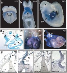
          microRNAs are regulators of myriad cellular events, but evidence for a single microRNA that can efficiently differentiate multipotent cells into a specific lineage or regulate direct reprogramming of cells into an alternate cell fate has been elusive. Here, we show that miR-145 and miR-143 are co-transcribed in multipotent cardiac progenitors before becoming localized to smooth muscle cells, including neural crest stem cell–derived vascular smooth muscle cells. miR-145 and miR-143 were direct transcriptional targets of serum response factor, myocardin and Nkx2.5, and were downregulated in injured or atherosclerotic vessels containing proliferating, less differentiated smooth muscle cells. miR-145 was necessary for myocardin-induced reprogramming of adult fibroblasts into smooth muscle cells and sufficient to induce differentiation of multipotent neural crest stem cells into vascular smooth muscle. Furthermore, miR-145 and miR-143 cooperatively targeted a network of transcription factors, including Klf4, myocardin, and Elk-1 to promote differentiation and repress proliferation of smooth muscle cells. These findings demonstrate that miR-145 can direct the smooth muscle fate and that miR-145 and miR-143 function to regulate the quiescent versus proliferative phenotype of smooth muscle cells.

          Related collections

          Most cited references24

          • Record: found
          • Abstract: found
          • Article: not found

          Switching from repression to activation: microRNAs can up-regulate translation.

          AU-rich elements (AREs) and microRNA target sites are conserved sequences in messenger RNA (mRNA) 3' untranslated regions (3'UTRs) that control gene expression posttranscriptionally. Upon cell cycle arrest, the ARE in tumor necrosis factor-alpha (TNFalpha) mRNA is transformed into a translation activation signal, recruiting Argonaute (AGO) and fragile X mental retardation-related protein 1 (FXR1), factors associated with micro-ribonucleoproteins (microRNPs). We show that human microRNA miR369-3 directs association of these proteins with the AREs to activate translation. Furthermore, we document that two well-studied microRNAs-Let-7 and the synthetic microRNA miRcxcr4-likewise induce translation up-regulation of target mRNAs on cell cycle arrest, yet they repress translation in proliferating cells. Thus, activation is a common function of microRNPs on cell cycle arrest. We propose that translation regulation by microRNPs oscillates between repression and activation during the cell cycle.
            Bookmark
            • Record: found
            • Abstract: found
            • Article: not found

            Serum response factor regulates a muscle-specific microRNA that targets Hand2 during cardiogenesis.

            Gradients of signalling and transcription factors govern many aspects of embryogenesis, highlighting the need for spatiotemporal control of regulatory protein levels. MicroRNAs are phylogenetically conserved small RNAs that regulate the translation of target messenger RNAs, providing a mechanism for protein dose regulation. Here we show that microRNA-1-1 (miR-1-1) and miR-1-2 are specifically expressed in cardiac and skeletal muscle precursor cells. We found that the miR-1 genes are direct transcriptional targets of muscle differentiation regulators including serum response factor, MyoD and Mef2. Correspondingly, excess miR-1 in the developing heart leads to a decreased pool of proliferating ventricular cardiomyocytes. Using a new algorithm for microRNA target identification that incorporates features of RNA structure and target accessibility, we show that Hand2, a transcription factor that promotes ventricular cardiomyocyte expansion, is a target of miR-1. This work suggests that miR-1 genes titrate the effects of critical cardiac regulatory proteins to control the balance between differentiation and proliferation during cardiogenesis.
              Bookmark
              • Record: found
              • Abstract: found
              • Article: not found

              Dysregulation of cardiogenesis, cardiac conduction, and cell cycle in mice lacking miRNA-1-2.

              MicroRNAs (miRNAs) are genomically encoded small RNAs used by organisms to regulate the expression of proteins generated from messenger RNA transcripts. The in vivo requirement of specific miRNAs in mammals through targeted deletion remains unknown, and reliable prediction of mRNA targets is still problematic. Here, we show that miRNA biogenesis in the mouse heart is essential for cardiogenesis. Furthermore, targeted deletion of the muscle-specific miRNA, miR-1-2, revealed numerous functions in the heart, including regulation of cardiac morphogenesis, electrical conduction, and cell-cycle control. Analyses of miR-1 complementary sequences in mRNAs upregulated upon miR-1-2 deletion revealed an enrichment of miR-1 "seed matches" and a strong tendency for potential miR-1 binding sites to be located in physically accessible regions. These findings indicate that subtle alteration of miRNA dosage can have profound consequences in mammals and demonstrate the utility of mammalian loss-of-function models in revealing physiologic miRNA targets.
                Bookmark

                Author and article information

                Journal
                0410462
                6011
                Nature
                Nature
                0028-0836
                1476-4687
                8 July 2009
                5 July 2009
                6 August 2009
                7 February 2010
                : 460
                : 7256
                : 705-710
                Affiliations
                [1 ]Gladstone Institute of Cardiovascular Disease, San Francisco, CA 94158, USA
                [2 ]Department of Pediatrics, University of California, San Francisco, CA 94543
                [3 ]Department of Biochemistry & Biophysics, University of California, San Francisco, CA 94143, USA
                [4 ]Aab Cardiovascular Research Institute, University of Rochester School of Medicine and Dentistry, Rochester, New York, 14642, USA
                Author notes
                [* ]Corresponding Author: dsrivastava@ 123456gladstone.ucsf.edu , Tel: (415) 734-2716, Fax: (415) 355-0141
                Article
                nihpa123667
                10.1038/nature08195
                2769203
                19578358
                2aa83001-850b-444b-884d-3bc643ae6d17
                History
                Funding
                Funded by: National Heart, Lung, and Blood Institute : NHLBI
                Award ID: R01 HL091168-01A1 ||HL
                Funded by: National Heart, Lung, and Blood Institute : NHLBI
                Award ID: R01 HL062572-12 ||HL
                Categories
                Article

                Uncategorized
                Uncategorized

                Comments

                Comment on this article