20
views
0
recommends
+1 Recommend
0 collections
    0
    shares
      • Record: found
      • Abstract: found
      • Article: found
      Is Open Access

      Custom-made 3D printed subperiosteal titanium implants for the prosthetic restoration of the atrophic posterior mandible of elderly patients: a case series

      research-article

      Read this article at

      Bookmark
          There is no author summary for this article yet. Authors can add summaries to their articles on ScienceOpen to make them more accessible to a non-specialist audience.

          Abstract

          Purpose

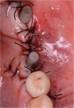
          To present the application of custom-made 3D-printed subperiosteal implants for fixed prosthetic restoration of the atrophic posterior mandible of elderly patients.

          Methods

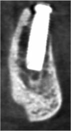
          Between January 2017 and June 2018, all partially edentulous patients aged over 65 years, with two or more missing teeth in the posterior atrophic mandible, and who did not want to undergo bone regenerative procedures, were included in this study. These patients were rehabilitated with custom-made subperiosteal implants, designed from cone beam computed tomography (CBCT) and fabricated in titanium by means of direct metal laser sintering (DMLS). The outcome measures were fit and stability of the implants at placement, duration of the intervention, implant survival, and early and late complications. All patients were followed for 1 year after surgery.

          Results

          Ten patients (four males, six females; mean age 69.6, SD ± 2.8, median 69, 95% CI 67.9–71.6) were included in the study. The fit of the implants was satisfactory, with a mean rating of 7 out of 10 (SD ± 1.6, median 7, 95% CI 6–8). Only two implants had insufficient fit, because of the presence of scattering in the CBCT; however, they were adapted to the sites during the interventions. The mean duration of the intervention was 44.3 min (SD ± 19.4, median 37, 95% CI 32.3–56.3). At the one-year follow-up, no implants were lost (survival rate 100%). One implant presented immediate postoperative complications with pain, discomfort and swelling, and two patients experienced late complications, having their provisional restorations fractured during the temporisation phase. All these complications were minor in nature, but the final complication rate amounted to 30% (three of ten patients).

          Conclusions

          Although this study has limits (small patient sample and short follow-up), DMLS has proven to be an effective method for fabricating accurate subperiosteal implants, with high survival rates. This may represent an alternative treatment procedure in elderly patients with a severely atrophic posterior mandible, since it allows avoidance of regenerative bone therapies. Further studies are needed to confirm these outcomes.

          Related collections

          Most cited references52

          • Record: found
          • Abstract: found
          • Article: found
          Is Open Access

          The complete digital workflow in fixed prosthodontics: a systematic review

          Background The continuous development in dental processing ensures new opportunities in the field of fixed prosthodontics in a complete virtual environment without any physical model situations. The aim was to compare fully digitalized workflows to conventional and/or mixed analog-digital workflows for the treatment with tooth-borne or implant-supported fixed reconstructions. Methods A PICO strategy was executed using an electronic (MEDLINE, EMBASE, Google Scholar) plus manual search up to 2016–09-16 focusing on RCTs investigating complete digital workflows in fixed prosthodontics with regard to economics or esthetics or patient-centered outcomes with or without follow-up or survival/success rate analysis as well as complication assessment of at least 1 year under function. The search strategy was assembled from MeSH-Terms and unspecific free-text words: {((“Dental Prosthesis” [MeSH]) OR (“Crowns” [MeSH]) OR (“Dental Prosthesis, Implant-Supported” [MeSH])) OR ((crown) OR (fixed dental prosthesis) OR (fixed reconstruction) OR (dental bridge) OR (implant crown) OR (implant prosthesis) OR (implant restoration) OR (implant reconstruction))} AND {(“Computer-Aided Design” [MeSH]) OR ((digital workflow) OR (digital technology) OR (computerized dentistry) OR (intraoral scan) OR (digital impression) OR (scanbody) OR (virtual design) OR (digital design) OR (cad/cam) OR (rapid prototyping) OR (monolithic) OR (full-contour))} AND {(“Dental Technology” [MeSH) OR ((conventional workflow) OR (lost-wax-technique) OR (porcelain-fused-to-metal) OR (PFM) OR (implant impression) OR (hand-layering) OR (veneering) OR (framework))} AND {((“Study, Feasibility” [MeSH]) OR (“Survival” [MeSH]) OR (“Success” [MeSH]) OR (“Economics” [MeSH]) OR (“Costs, Cost Analysis” [MeSH]) OR (“Esthetics, Dental” [MeSH]) OR (“Patient Satisfaction” [MeSH])) OR ((feasibility) OR (efficiency) OR (patient-centered outcome))}. Assessment of risk of bias in selected studies was done at a ‘trial level’ including random sequence generation, allocation concealment, blinding, completeness of outcome data, selective reporting, and other bias using the Cochrane Collaboration tool. A judgment of risk of bias was assigned if one or more key domains had a high or unclear risk of bias. An official registration of the systematic review was not performed. Results The systematic search identified 67 titles, 32 abstracts thereof were screened, and subsequently, three full-texts included for data extraction. Analysed RCTs were heterogeneous without follow-up. One study demonstrated that fully digitally produced dental crowns revealed the feasibility of the process itself; however, the marginal precision was lower for lithium disilicate (LS2) restorations (113.8 μm) compared to conventional metal-ceramic (92.4 μm) and zirconium dioxide (ZrO2) crowns (68.5 μm) (p < 0.05). Another study showed that leucite-reinforced glass ceramic crowns were esthetically favoured by the patients (8/2 crowns) and clinicians (7/3 crowns) (p < 0.05). The third study investigated implant crowns. The complete digital workflow was more than twofold faster (75.3 min) in comparison to the mixed analog-digital workflow (156.6 min) (p < 0.05). No RCTs could be found investigating multi-unit fixed dental prostheses (FDP). Conclusions The number of RCTs testing complete digital workflows in fixed prosthodontics is low. Scientifically proven recommendations for clinical routine cannot be given at this time. Research with high-quality trials seems to be slower than the industrial progress of available digital applications. Future research with well-designed RCTs including follow-up observation is compellingly necessary in the field of complete digital processing. Electronic supplementary material The online version of this article (10.1186/s12903-017-0415-0) contains supplementary material, which is available to authorized users.
            Bookmark
            • Record: found
            • Abstract: found
            • Article: found
            Is Open Access

            Cone beam computed tomography in implant dentistry: recommendations for clinical use

            Background In implant dentistry, three-dimensional (3D) imaging can be realised by dental cone beam computed tomography (CBCT), offering volumetric data on jaw bones and teeth with relatively low radiation doses and costs. The latter may explain why the market has been steadily growing since the first dental CBCT system appeared two decades ago. More than 85 different CBCT devices are currently available and this exponential growth has created a gap between scientific evidence and existing CBCT machines. Indeed, research for one CBCT machine cannot be automatically applied to other systems. Methods Supported by a narrative review, recommendations for justified and optimized CBCT imaging in oral implant dentistry are provided. Results The huge range in dose and diagnostic image quality requires further optimization and justification prior to clinical use. Yet, indications in implant dentistry may go beyond diagnostics. In fact, the inherent 3D datasets may further allow surgical planning and transfer to surgery via 3D printing or navigation. Nonetheless, effective radiation doses of distinct dental CBCT machines and protocols may largely vary with equivalent doses ranging between 2 to 200 panoramic radiographs, even for similar indications. Likewise, such variation is also noticed for diagnostic image quality, which reveals a massive variability amongst CBCT technologies and exposure protocols. For anatomical model making, the so-called segmentation accuracy may reach up to 200 μm, but considering wide variations in machine performance, larger inaccuracies may apply. This also holds true for linear measures, with accuracies of 200 μm being feasible, while sometimes fivefold inaccuracy levels may be reached. Diagnostic image quality may also be dramatically hampered by patient factors, such as motion and metal artefacts. Apart from radiodiagnostic possibilities, CBCT may offer a huge therapeutic potential, related to surgical guides and further prosthetic rehabilitation. Those additional opportunities may surely clarify part of the success of using CBCT for presurgical implant planning and its transfer to surgery and prosthetic solutions. Conclusions Hence, dental CBCT could be justified for presurgical diagnosis, preoperative planning and peroperative transfer for oral implant rehabilitation, whilst striving for optimisation of CBCT based machine-dependent, patient-specific and indication-oriented variables.
              Bookmark
              • Record: found
              • Abstract: found
              • Article: not found

              Clinical outcomes of vertical bone augmentation to enable dental implant placement: a systematic review.

              This review addressed the focused question of what is the predictability of vertical ridge augmentation techniques for patients, who were diagnosed with insufficient alveolar bone volume for the placement of dental implants.
                Bookmark

                Author and article information

                Contributors
                camangan@gmail.com
                andrea.ebianchi@gmail.com
                francescomangano1@mclink.net
                jessica.dana@bluewin.ch
                m.colombo.dds@gmail.com
                solopivan@yandex.ru
                admakin1966@mail.ru
                Journal
                3D Print Med
                3D Print Med
                3D Printing in Medicine
                Springer International Publishing (Cham )
                2365-6271
                8 January 2020
                8 January 2020
                December 2020
                : 6
                : 1
                Affiliations
                [1 ]GRID grid.15496.3f, Department of Dental Sciences, , University Vita Salute S. Raffaele, ; 20132 Milan, Italy
                [2 ]Department of Periodontology and Implantology, Istituto Stomatologico Italiano, 20122 Milan, Italy
                [3 ]ISNI 0000 0001 2288 8774, GRID grid.448878.f, Department of Prevention and Communal Dentistry, , Sechenov First Moscow State Medical University, ; 119991 Moscow, Russia
                [4 ]Private Practice, 6918 Lugano, Switzerland
                [5 ]Private Practice, 21100 Varese, Italy
                Article
                55
                10.1186/s41205-019-0055-x
                6950914
                31915946
                2b0ced66-51fe-43bc-bd46-4d633bb4bd27
                © The Author(s) 2020

                Open AccessThis article is distributed under the terms of the Creative Commons Attribution 4.0 International License ( http://creativecommons.org/licenses/by/4.0/), which permits unrestricted use, distribution, and reproduction in any medium, provided you give appropriate credit to the original author(s) and the source, provide a link to the Creative Commons license, and indicate if changes were made.

                History
                : 23 September 2019
                : 20 December 2019
                Categories
                Research
                Custom metadata
                © The Author(s) 2020

                custom-made,subperiosteal implants,direct metal laser sintering (dmls),atrophic posterior mandible,elderly patients

                Comments

                Comment on this article