Inviting an author to review:
Find an author and click ‘Invite to review selected article’ near their name.
Search for authorsSearch for similar articles
11
views
0
recommends
+1 Recommend
0 collections
    0
    shares
      • Record: found
      • Abstract: found
      • Article: found
      Is Open Access

      Laser-induced vapour nanobubbles improve drug diffusion and efficiency in bacterial biofilms

      research-article

      Read this article at

      Bookmark
          There is no author summary for this article yet. Authors can add summaries to their articles on ScienceOpen to make them more accessible to a non-specialist audience.

          Abstract

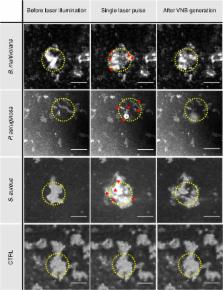
          Hindered penetration of antibiotics through biofilms is one of the reasons for the alarming increase in bacterial tolerance to antibiotics. Here, we investigate the potential of laser-induced vapour nanobubbles (VNBs) formed around plasmonic nanoparticles to locally disturb biofilm integrity and improve antibiotics diffusion. Our results show that biofilms of both Gram-negative ( Burkholderia multivorans, Pseudomonas aeruginosa) and Gram-positive ( Staphylococcus aureus) bacteria can be loaded with cationic 70-nm gold nanoparticles and that subsequent laser illumination results in VNB formation inside the biofilms. In all types of biofilms tested, VNB formation leads to substantial local biofilm disruption, increasing tobramycin efficacy up to 1-3 orders of magnitude depending on the organism and treatment conditions. Altogether, our results support the potential of laser-induced VNBs as a new approach to disrupt biofilms of a broad range of organisms, resulting in improved antibiotic diffusion and more effective biofilm eradication.

          Abstract

          Eradication of bacterial infections can be hindered by poor penetration of antibiotics through biofilms. Here, Teirlinck et al. show that laser-induced vapour nanobubbles formed around plasmonic nanoparticles can be used to locally disturb biofilm integrity and improve antibiotic diffusion.

          Related collections

          Most cited references64

          • Record: found
          • Abstract: found
          • Article: not found

          Caenorhabditis elegans: An Emerging Model in Biomedical and Environmental Toxicology

          The nematode Caenorhabditis elegans has emerged as an important animal model in various fields including neurobiology, developmental biology, and genetics. Characteristics of this animal model that have contributed to its success include its genetic manipulability, invariant and fully described developmental program, well-characterized genome, ease of maintenance, short and prolific life cycle, and small body size. These same features have led to an increasing use of C. elegans in toxicology, both for mechanistic studies and high-throughput screening approaches. We describe some of the research that has been carried out in the areas of neurotoxicology, genetic toxicology, and environmental toxicology, as well as high-throughput experiments with C. elegans including genome-wide screening for molecular targets of toxicity and rapid toxicity assessment for new chemicals. We argue for an increased role for C. elegans in complementing other model systems in toxicological research.
            Bookmark
            • Record: found
            • Abstract: found
            • Article: not found

            Biofilm dispersal: mechanisms, clinical implications, and potential therapeutic uses.

            J Kaplan (2010)
            Like all sessile organisms, surface-attached communities of bacteria known as biofilms must release and disperse cells into the environment to colonize new sites. For many pathogenic bacteria, biofilm dispersal plays an important role in the transmission of bacteria from environmental reservoirs to human hosts, in horizontal and vertical cross-host transmission, and in the exacerbation and spread of infection within a host. The molecular mechanisms of bacterial biofilm dispersal are only beginning to be elucidated. Biofilm dispersal is a promising area of research that may lead to the development of novel agents that inhibit biofilm formation or promote biofilm cell detachment. Such agents may be useful for the prevention and treatment of biofilms in a variety of industrial and clinical settings. This review describes the current status of research on biofilm dispersal, with an emphasis on studies aimed to characterize dispersal mechanisms, and to identify environmental cues and inter- and intracellular signals that regulate the dispersal process. The clinical implications of biofilm dispersal and the potential therapeutic applications of some of the most recent findings will also be discussed.
              Bookmark
              • Record: found
              • Abstract: not found
              • Article: not found

              Recent biomedical applications of gold nanoparticles: A review

                Bookmark

                Author and article information

                Contributors
                Kevin.Braeckmans@UGent.be
                Journal
                Nat Commun
                Nat Commun
                Nature Communications
                Nature Publishing Group UK (London )
                2041-1723
                30 October 2018
                30 October 2018
                2018
                : 9
                : 4518
                Affiliations
                [1 ]ISNI 0000 0001 2069 7798, GRID grid.5342.0, Laboratory of General Biochemistry and Physical Pharmacy, , University of Ghent, ; Ghent, 9000 Belgium
                [2 ]Centre for Nano- and Biophotonics, Ghent, 9000 Belgium
                [3 ]ISNI 0000 0004 0626 3303, GRID grid.410566.0, Laboratory of Toxicology, , Ghent University Hospital, ; Ghent, 9000 Belgium
                [4 ]ISNI 0000 0001 2069 7798, GRID grid.5342.0, Laboratory of Pharmaceutical Microbiology, , University of Ghent, ; Ghent, 9000 Belgium
                [5 ]ISNI 0000 0001 2069 7798, GRID grid.5342.0, Department of Biomedical Molecular Biology, VIB Center for Inflammation Research, , Ghent University, ; 9052 Ghent, Belgium
                [6 ]ISNI 0000 0001 2069 7798, GRID grid.5342.0, Expertise Centre for Transmission Electron Microscopy, VIB BioImaging Core, , Ghent University, ; Ghent, 9052 Belgium
                [7 ]ISNI 0000 0004 0638 7509, GRID grid.464109.e, IEMN UMR 8520, , Université de Lille, ; Villeneuve d’Ascq, 59652 France
                [8 ]ISNI 0000 0004 0368 4014, GRID grid.462765.4, Laboratoire de Physique des Lasers, Atomes et Molécules UMR 8523, ; Villeneuve d’Ascq, 59655 France
                Author information
                http://orcid.org/0000-0002-6518-3835
                http://orcid.org/0000-0001-7386-7164
                http://orcid.org/0000-0002-4887-2161
                http://orcid.org/0000-0002-7145-9826
                http://orcid.org/0000-0002-6407-0601
                http://orcid.org/0000-0002-7993-6295
                Article
                6884
                10.1038/s41467-018-06884-w
                6207769
                30375378
                2cd1aac3-f62c-462b-be30-e01cf42d1a03
                © The Author(s) 2018

                Open Access This article is licensed under a Creative Commons Attribution 4.0 International License, which permits use, sharing, adaptation, distribution and reproduction in any medium or format, as long as you give appropriate credit to the original author(s) and the source, provide a link to the Creative Commons license, and indicate if changes were made. The images or other third party material in this article are included in the article’s Creative Commons license, unless indicated otherwise in a credit line to the material. If material is not included in the article’s Creative Commons license and your intended use is not permitted by statutory regulation or exceeds the permitted use, you will need to obtain permission directly from the copyright holder. To view a copy of this license, visit http://creativecommons.org/licenses/by/4.0/.

                History
                : 31 January 2018
                : 2 October 2018
                Funding
                Funded by: FundRef https://doi.org/10.13039/501100003130, Fonds Wetenschappelijk Onderzoek (Research Foundation Flanders);
                Award ID: 1500418N
                Award ID: 1500418N
                Award Recipient :
                Funded by: FundRef https://doi.org/10.13039/501100003132, Agentschap voor Innovatie door Wetenschap en Technologie (Agency for Innovation by Science and Technology, Flanders);
                Award ID: 01G02215
                Award Recipient :
                Funded by: FundRef https://doi.org/10.13039/501100000781, EC | European Research Council (ERC);
                Award ID: 648124
                Award Recipient :
                Categories
                Article
                Custom metadata
                © The Author(s) 2018

                Uncategorized
                Uncategorized

                Comments

                Comment on this article