- Record: found
- Abstract: found
- Article: found
Focal Dermal Hypoplasia or Goltz Syndrome: A Rare Association with Keratoconus
letter
Read this article at
There is no author summary for this article yet. Authors can add summaries to their articles on ScienceOpen to make them more accessible to a non-specialist audience.
Abstract
Sir,
Focal dermal hypoplasia (FDH) or Goltz syndrome was first described by Liebermann
in 1935 as “atrophodermia linearis maculosa et papillomatosis congenitalis.” Goltz
in 1962 mentioned the term “FDH.” It is a rare syndrome identified by dysplasia of
structures derived from ectoderm and mesoderm. Hence, named as congenital ectodermal-mesodermal
dysplasia also.[1] Incidence is 1:50,000–1:150,000 in the general population. It primarily
affects the cutaneous and skeletal system, but ocular, dental, and central nervous
system anomalies are fairly common. Being an X-linked dominant condition only females
are affected, whereas male dies in utero. Skeletal abnormalities are found in around
80% cases.[2]
We report a case of FDH in an 11 weeks, low birth weight female infant born out of
nonconsanguineous marriage from an unaffected parent.
At the time of presentation to us she had multiple inflamed and eroded patches over
posterior thigh, back, and buttock [Figure 1]. After 6 weeks, she developed multiple
round hypopigmented atrophied patches of varying sizes developed bilaterally over
the back of thigh, buttock, lateral abdomen, and umbilicus in a blaschkoid pattern.
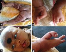
Lesions were cribriform and surrounded by a hyperpigmented border [Figure 2a]. On
skeletal examination, there were some typical changes. Hand feet showed syndactyly
of right 3rd and 4th finger, polydactyly of left foot, and lobster deformity of the
right foot [Figure 2b and d]. Nails of hand were dystrophic. Face showed round shape,
pointed chin, incomplete left-sided cleft lip, narrow depressed nasal bridge, widened
flared nasal ala with less scalp, and eyebrow hair [Figure 2c]. On ophthalmology referral,
they found bilateral keratoconus. X-ray from long bones showed osteopathia striata.
Biopsy from the atrophic, hypopigmented lesion of the back of thigh showed normal
epidermis and thin dermis with scanty collagen. Multiple discrete areas of mature
adipocytes impinging epidermis noted. This rare skin disease is due to a mutation
in PORCN gene that is located in X chromosome. PORCN is a part of porcupine gene family
detected first in drosophila. It is responsible for the synthesis of endoplasmic reticulum
protein with multiple transmembrane domains. It is also a regulator of Wnt signaling.[3]
Eye features include microphthalmia with bilateral coloboma of the iris, ectopia lentis,
strabismus, anophthalmia, nystagmus, irregularities of the pupils, and corneal defects
such as keratoconus, photophobia, and ptosis.[4] Eye lesions itself are not so common
with this syndrome and keratoconus was reported in very few cases. Musculoskeletal
defects are osteopenia, spina bifida, scoliosis, etc. Hypermobile joint with hand
foot asymmetry is evident many times. Syndactyly, ectrodactyly polydactyly, hypoplasia,
or agenesis of fingers with a lobster deformity in extreme cases is seen among hand
foot defect.[5] The lobster defect of the foot was seen in our cases also.
Figure 1
Inflamed eroded areas at presentation
Figure 2
(a) Cribriform atrophy over posterior thigh (b) lobster foot (c) left-sided partial
cleft lip (d) lobster foot(closer view)
Treatment of Goltz syndrome is mainly supportive. Genetic counseling, local skin care,
correction of systemic associations and deformities are the mainstay of therapy. Proper
rehabilitative measures are of utmost importance for a grown up child.[4]
Declaration of patient consent
The authors certify that they have obtained all appropriate patient consent forms.
In the form the patient(s) has/have given his/her/their consent for his/her/their
images and other clinical information to be reported in the journal. The patients
understand that their names and initials will not be published and due efforts will
be made to conceal their identity, but anonymity cannot be guaranteed.
Financial support and sponsorship
Nil.
Conflicts of interest
There are no conflicts of interest.
Related collections
Most cited references5
- Record: found
- Abstract: found
- Article: not found
Deficiency of PORCN, a regulator of Wnt signaling, is associated with focal dermal hypoplasia.
Mauro Paradisi, Rudolf Happle, Barbara Fritz … (2007)
- Record: found
- Abstract: not found
- Article: not found
Focal dermal hypoplasia.
R GOLTZ, R Gorlin, H G RAVITS … (1962)

- Record: found
- Abstract: found
- Article: found
Goltz syndrome (focal dermal hypoplasia) with unilateral ocular, cutaneous and skeletal features: case report
Addis Tenkir, Samuel Teshome (2010)