13
views
0
recommends
+1 Recommend
0 collections
    0
    shares
      • Record: found
      • Abstract: found
      • Article: found
      Is Open Access

      Internal jugular vein duplication

      letter

      Read this article at

      Bookmark
          There is no author summary for this article yet. Authors can add summaries to their articles on ScienceOpen to make them more accessible to a non-specialist audience.

          Abstract

          Sir, The internal jugular vein is the largest vein in the neck and drains the intracranial structures and deep structures of the face and neck. It runs the length of the neck slightly lateral to the common carotid artery within the carotid sheath and, on the right, crosses in front of the right subclavian artery, to join the subclavian vein.[1] Duplication of the internal jugular vein is a rare finding. We came across a case of duplication of right internal jugular vein and offer some clinical comments on the importance of this rare anatomical feature. A 65-year-old woman with a T2N0M0 squamous cell carcinoma of the right margin of the tongue had a local wide excision and an ipsilateral modified type 3 radical neck dissection. 
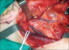
During the dissection of the neck a bifurcation of the right internal jugular vein, about 2cm from the jugular foramen was encountered [Figure 1]. Both branches of the internal jugular vein had the same thickness and poured into the right subclavian vein. The anterior branch was parallel to the carotid artery and received the common facial vein, the superior and inferior thyroid veins and the transverse cervical vein. The posterior branch passed within the carotid sheath, drained only the cerebral blood and emptied into the subclavian vein lateral to the medial branch. None showed evidence of phlebectasia or aneurysm. The spinal accessory nerve passed between the medial and lateral branches, exactly superficial to the medial branch and under the lateral branch. Figure 1 The bifurcation of the right internal jugular vein discovered during neck dissection Duplication of the internal jugular vein is a rare congenital anomaly. The vein divides into two branches that separately enter the subclavian vein. Almost always it involves the upper third of the vein.[2 3] In our case, the duplication was 2 cm below the base of the skull. Duplication of the internal jugular vein is usually reported in association with phlebectasia, which is a soft non-pulsatile cervical swelling that increases in size during Valsalva maneuver.[4] In our case, no aneurysm or phlebectasia was observed. Three theories have been formulated to explain duplication:[3] the vascular theory, that is usually accepted[3] the neural hypothesis and the bony hypothesis. Duplication is thought to result from the appearance of a secondary venous ring at a lower level surrounding the spinal accessory nerve during foetal life.[4] The persistence of this secondary ring in adult life may be important in the aetiology of venous duplication.[4] Unexpected duplication of the internal jugular vein could impact specific clinical procedures, creating the possibility of either iatrogenic morbidity or incorrect diagnosis. In our case, the large area involved and additional sites for ligature complicated selective neck dissection.

          Related collections

          Most cited references4

          • Record: found
          • Abstract: found
          • Article: not found

          High duplication of the internal jugular vein: clinical incidence in the adult and surgical consequences, a report of three clinical cases.

          Duplication of the internal jugular vein (IJV) is a rare malformation. Three intraoperative cases are reported. In our personal experience, the clinical incidence of the anomaly is approximately 4 per 1,000 unilateral neck dissections. The venous duplication is at a variable height, affecting the superior part of the IJV. The lateral branch of the accessory nerve (XI) always passes medially to the anterior vein and laterally to the posterior vein, between the venous duplication. This is most often unilateral but sometimes bilateral. The IJV may be normal, dilated or ectatic. The discovery of this anatomical variation has practical implications during cervical lymph node clearance, either functional or radical, during oncological surgery necessitating viewing the IJV and its affluents and the lateral branch of the accessory nerve. The embryological explanation suggests a topographical "conflict" between the development of the IJV and the lateral branch of the accessory nerve. The French version of this article is available in the form of electronic supplementary material and can be obtained by using the Springer LINK server located at http://dx.doi.org/10.1007/s00276-002-0020-y.
            Bookmark
            • Record: found
            • Abstract: found
            • Article: not found

            Bilateral duplicated internal jugular veins: case study and literature review.

            A rare bilateral duplication of the internal jugular vein (IJV) was discovered during cadaveric dissection. From each jugular foramen, a single IJV descended to the level of the hyoid bone then divided into medial and lateral veins. The medial IJVs traveled in the carotid sheath; the lateral IJVs coursed posterolateral to the sheath across the lateral cervical region (posterior triangle) of the neck. On the right side, medial and lateral IJVs entered the subclavian vein separately. C2-C3 anterior rami and the suprascapular artery passed between the medial and lateral IJVs. The right external jugular vein passed aberrantly between the heads of the sternocleidomastoid muscle (SCM) into the subclavian vein anterior to the lateral IJV. On the left side, the medial IJV drained into a large bulbous jugulovertebrosubclavian (JVS) sinus that received six main vessels. The lateral IJV diverged posterolaterally toward the border of the trapezius muscle, received the transverse cervical vein, and then turned sharply anteromedially to drain into the JVS sinus. The lateral IJV also gave an aberrant additional large vein that passed laterally around the omohyoid muscle before entering the JVS sinus. The left external jugular vein paralleled the anterior border of SCM before passing posterolaterally to terminate in the JVS sinus. Jugular vein anomalies of this magnitude are very rare. Determining the frequency of multiple IJVs is hampered by inconsistent terminology. We suggest that IJV duplication differs from fenestration anatomically and, potentially, developmentally. Criteria for characterizing IJV duplication and fenestration are proposed. The mechanism of development and the clinical significance of multiple IJVs are discussed.
              Bookmark
              • Record: found
              • Abstract: found
              • Article: not found

              Duplication of internal jugular vein and relation to the spinal accessory nerve.

              We report a case of duplication of the internal jugular vein in which the duplication was incomplete and the accessory nerve lay deep to the vein.
                Bookmark

                Author and article information

                Journal
                Indian J Plast Surg
                IJPS
                Indian Journal of Plastic Surgery : Official Publication of the Association of Plastic Surgeons of India
                Medknow Publications (India )
                0970-0358
                1998-376X
                Jul-Dec 2009
                : 42
                : 2
                : 273-274
                Affiliations
                Maxillo-Facial Surgery, Head and Neck Department, II University of Naples, Naples, Italy
                [1 ]Plastic Surgery, Rome, Italy
                Author notes
                Address for correspondence: Dr. Raffaele Rauso, Corso A. Moro, 100, 81055, S. Maria C. V. (CE), Italy. E-mail: raffaelerauso@ 123456virgilio.it
                Article
                IJPS-42-273
                10.4103/0970-0358.59303
                2845386
                20368879
                2e1baba1-ca01-4cba-8ce2-702f173470ed
                © Indian Journal of Plastic Surgery

                This is an open-access article distributed under the terms of the Creative Commons Attribution License, which permits unrestricted use, distribution, and reproduction in any medium, provided the original work is properly cited.

                History
                Categories
                Letters to Editor

                Surgery
                Surgery

                Comments

                Comment on this article