21
views
0
recommends
+1 Recommend
0 collections
    0
    shares
      • Record: found
      • Abstract: found
      • Article: found
      Is Open Access

      Shear-Wave Elastography Assessments of Quadriceps Stiffness Changes prior to, during and after Prolonged Exercise: A Longitudinal Study during an Extreme Mountain Ultra-Marathon

      research-article

      Read this article at

      Bookmark
          There is no author summary for this article yet. Authors can add summaries to their articles on ScienceOpen to make them more accessible to a non-specialist audience.

          Abstract

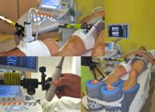
          In sports medicine, there is increasing interest in quantifying the elastic properties of skeletal muscle, especially during extreme muscular stimulation, to improve our understanding of the impact of alterations in skeletal muscle stiffness on resulting pain or injuries, as well as the mechanisms underlying the relationships between these parameters. Our main objective was to determine whether real-time shear-wave elastography (SWE) can monitor changes in quadriceps muscle elasticity during an extreme mountain ultra-marathon, a powerful mechanical stress model. Our study involved 50 volunteers participating in an extreme mountain marathon (distance: 330 km, elevation: +24,000 m). Quantitative SWE velocity and shear modulus measurements were performed in most superficial quadriceps muscle heads at the following 4 time points: before the race, halfway through the race, upon finishing the race and after recovery (+48 h). Blood biomarker levels were also measured. A significant decrease in the quadriceps shear modulus was observed upon finishing the race (3.31±0.61 kPa) (p<0.001) compared to baseline (3.56±0.63 kPa), followed by a partial recovery +48 h after the race (3.45±0.6 kPa) (p = 0.002) across all muscle heads, as well as for each of the following three muscle heads: the rectus femoris (p = 0.003), the vastus medialis (p = 0.033) and the vastus lateralis (p = 0.001). Our study is the first to assess changes in muscle stiffness during prolonged extreme physical endurance exercises based on shear modulus measurements using non-invasive SWE. We concluded that decreases in stiffness, which may have resulted from quadriceps overuse in the setting of supra-physiological stress caused by the extreme distance and unique elevation of the race, may have been responsible for the development of inflammation and muscle swelling. SWE may hence represent a promising tool for monitoring physiologic or pathological variations in muscle stiffness and may be useful for diagnosing and monitoring muscle changes.

          Related collections

          Most cited references57

          • Record: found
          • Abstract: found
          • Article: not found

          Muscle function after exercise-induced muscle damage and rapid adaptation.

          This brief review focuses on the time course of changes in muscle function and other correlates of muscle damage following maximal effort eccentric actions of the forearm flexor muscles. Data on 109 subjects are presented to describe an accurate time course of these changes and attempt to establish relationships among the measures. Peak soreness is experienced 2-3 d postexercise while peak swelling occurs 5 d postexercise. Maximal strength and the ability to fully flex the arm show the greatest decrements immediately after exercise with a linear restoration of these functions over the next 10 d. Blood creatine kinase (CK) levels increase precipitously at 2 d after exercise which is also the time when spontaneous muscle shortening is most pronounced. Whether the similarity in the time courses of some of these responses implies that they are caused by similar factors remains to be determined. Performance of one bout of eccentric exercise produces an adaptation such that the muscle is more resistant to damage from a subsequent bout of exercise. The length of the adaptation differs among the measures such that when the exercise regimens are separated by 6 wk, all measures show a reduction in response on the second, compared with the first, bout. After 10 wk, only CK and muscle shortening show a reduction in response. After 6 months only the CK response is reduced. A combination of cellular factors and neurological factors may be involved in the adaptation process.
            Bookmark
            • Record: found
            • Abstract: found
            • Article: not found

            Viscoelastic and anisotropic mechanical properties of in vivo muscle tissue assessed by supersonic shear imaging.

            The in vivo assessment of the biomechanical properties of the skeletal muscle is a complex issue because the muscle is an anisotropic, viscoelastic and dynamic medium. In this article, these mechanical properties are characterized for the brachialis muscle in vivo using a noninvasive ultrasound-based technique. This supersonic shear imaging technique combines an ultra-fast ultrasonic system and the remote generation of transient mechanical forces into tissue via the radiation force of focused ultrasonic beams. Such an ultrasonic radiation force is induced deep within the muscle by a conventional ultrasonic probe and the resulting shear waves are then imaged with the same probe (5 MHz) at an ultra-fast framerate (up to 5000 frames/s). Local tissue velocity maps are obtained with a conventional speckle tracking technique and provide a full movie of the shear wave propagation through the entire muscle. Shear wave group velocities are then estimated using a time of flight algorithm. This approach provides a complete set of quantitative and in vivo parameters describing the muscle's mechanical properties as a function of active voluntary contraction as well as passive extension of healthy volunteers. Anisotropic properties are also estimated by tilting the probe head with respects to the main muscular fibers direction. Finally, the dispersion of the shear waves is studied for these different configurations and shear modulus and shear viscosity are quantitatively assessed assuming the viscoelastic Voigt's model. Copyright 2010 World Federation for Ultrasound in Medicine & Biology. Published by Elsevier Inc. All rights reserved.
              Bookmark
              • Record: found
              • Abstract: found
              • Article: not found

              Muscle damage from eccentric exercise: mechanism, mechanical signs, adaptation and clinical applications.

              In eccentric exercise the contracting muscle is forcibly lengthened; in concentric exercise it shortens. While concentric contractions initiate movements, eccentric contractions slow or stop them. A unique feature of eccentric exercise is that untrained subjects become stiff and sore the day afterwards because of damage to muscle fibres. This review considers two possible initial events as responsible for the subsequent damage, damage to the excitation-contraction coupling system and disruption at the level of the sarcomeres. Other changes seen after eccentric exercise, a fall in active tension, shift in optimum length for active tension, and rise in passive tension, are seen, on balance, to favour sarcomere disruption as the starting point for the damage. As well as damage to muscle fibres there is evidence of disturbance of muscle sense organs and of proprioception. A second period of exercise, a week after the first, produces much less damage. This is the result of an adaptation process. One proposed mechanism for the adaptation is an increase in sarcomere number in muscle fibres. This leads to a secondary shift in the muscle's optimum length for active tension. The ability of muscle to rapidly adapt following the damage from eccentric exercise raises the possibility of clinical applications of mild eccentric exercise, such as for protecting a muscle against more major injuries.
                Bookmark

                Author and article information

                Contributors
                Role: Editor
                Journal
                PLoS One
                PLoS ONE
                plos
                plosone
                PLoS ONE
                Public Library of Science (San Francisco, CA USA )
                1932-6203
                31 August 2016
                2016
                : 11
                : 8
                : e0161855
                Affiliations
                [1 ]Department of Radiology, CHU Saint-Etienne, Université Jean-Monnet, Saint-Étienne, France
                [2 ]Univ Lyon, UJM-Saint-Etienne, INSA, CNRS UMR 5520, INSERM U1206, CREATIS, F-42023, Saint-Etienne, France
                [3 ]EuSpLM, Department of Clinical Chemistry, University of Liège, CHU Sart-Tilman, Liège, Belgium
                [4 ]Department of Intensive Care, CHU Saint-Etienne, Saint-Etienne, France
                [5 ]Department of Neurology and Neurophysiology, Mountain Medicine Center, Valle d’Aosta Regional Hospital, Aosta, Italy
                [6 ]Institute of Sport Sciences of the University of Lausanne (ISSUL), Lausanne, Switzerland
                Universite de Nantes, FRANCE
                Author notes

                Competing Interests: Supersonic Imagine contributed to this study by providing two of their ultrasound diagnostic systems equipped with the most advanced SWE module during the Tor des Geants. The authors also thank Sysmex for their support in providing a pocH-100i™ automated hematology analyzer (Sysmex, Villepinte, France). There are no patents, products in development or marketed products to declare. This does not alter our adherence to all the PLOS ONE policies on sharing data and materials. These industrial partners had no role in study design, data collection and analysis, decision to publish, or preparation of the manuscript.

                • Conceptualization: MV PC LG GM ClG.

                • Data curation: MV PC.

                • Formal analysis: PA PC MV.

                • Funding acquisition: MV PC.

                • Investigation: PA MV ClG CdB CT JM PC.

                • Methodology: MV PC.

                • Project administration: MV PC GG GM ClG LG.

                • Resources: MV PC GM GG.

                • Supervision: PC MV.

                • Validation: PA CdB CT MV PC.

                • Visualization: MV PA PC.

                • Writing – original draft: PA MV PC.

                • Writing – review & editing: ClG GM LG MV PA PC.

                Article
                PONE-D-16-07667
                10.1371/journal.pone.0161855
                5007013
                27579699
                2e5efcff-4cf9-4bbe-b48a-f307c7a0eb16
                © 2016 Andonian et al

                This is an open access article distributed under the terms of the Creative Commons Attribution License, which permits unrestricted use, distribution, and reproduction in any medium, provided the original author and source are credited.

                History
                : 23 February 2016
                : 13 August 2016
                Page count
                Figures: 4, Tables: 5, Pages: 21
                Funding
                The authors are especially grateful to Supersonic Imagine, who contributed to this study by providing two of their ultrasound diagnostic systems equipped with the most advanced SWE module during the Tor des Geants. The authors also thank Sysmex for their support in providing a pocH-100i™ automated hematology analyzer (Sysmex, Villepinte, France). This work was performed within the framework of the LABEX PRIMES (ANR-11-LABX-0063) of Université de Lyon, within the program "Investissements d'Avenir" (ANR-11-IDEX-0007) operated by the French National Research Agency (ANR). The funders had no role in study design, data collection and analysis, decision to publish, or preparation of the manuscript.
                Categories
                Research Article
                Research and Analysis Methods
                Bioassays and Physiological Analysis
                Muscle Analysis
                Biology and Life Sciences
                Anatomy
                Musculoskeletal System
                Muscles
                Skeletal Muscles
                Medicine and Health Sciences
                Anatomy
                Musculoskeletal System
                Muscles
                Skeletal Muscles
                Biology and Life Sciences
                Physiology
                Muscle Physiology
                Muscle Contraction
                Medicine and Health Sciences
                Physiology
                Muscle Physiology
                Muscle Contraction
                Biology and Life Sciences
                Biochemistry
                Biomarkers
                Physical Sciences
                Materials Science
                Material Properties
                Mechanical Properties
                Stiffness
                Biology and Life Sciences
                Immunology
                Immune Response
                Inflammation
                Medicine and Health Sciences
                Immunology
                Immune Response
                Inflammation
                Medicine and Health Sciences
                Diagnostic Medicine
                Signs and Symptoms
                Inflammation
                Medicine and Health Sciences
                Pathology and Laboratory Medicine
                Signs and Symptoms
                Inflammation
                Medicine and Health Sciences
                Sports and Exercise Medicine
                Biology and Life Sciences
                Sports Science
                Sports and Exercise Medicine
                Medicine and Health Sciences
                Diagnostic Medicine
                Diagnostic Radiology
                Ultrasound Imaging
                Research and Analysis Methods
                Imaging Techniques
                Diagnostic Radiology
                Ultrasound Imaging
                Medicine and Health Sciences
                Radiology and Imaging
                Diagnostic Radiology
                Ultrasound Imaging
                Custom metadata
                The authors confirm that all Shear-Wave-Elastrography data underlying the findings are fully available without restriction. Data are made available on Open Science Framework data repository at osf.io/e9y6c/?view_only=524b4a1e7da24662b71e2da7cadf349b.

                Uncategorized
                Uncategorized

                Comments

                Comment on this article