1
views
0
recommends
+1 Recommend
0 collections
    0
    shares
      • Record: found
      • Abstract: found
      • Article: found
      Is Open Access

      Vitreous Hemorrhage as Presenting Sign of Retinal Arteriovenous Malformation

      case-report

      Read this article at

      Bookmark
          There is no author summary for this article yet. Authors can add summaries to their articles on ScienceOpen to make them more accessible to a non-specialist audience.

          Abstract

          Objective

          To describe a patient with vitreous hemorrhage and peripheral retinal ischemia, eventually diagnosed with an underlying retinal arteriovenous malformation.

          Methods

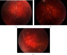
          A 15-year-old girl presented with sudden-onset, painless visual loss in the right eye. She underwent a full ophthalmological work-up.

          Results

          BCVA was less than 20/400 in the right eye and 20/20 in the left eye. Intraocular pressure and anterior segment examination were unremarkable. Fundoscopy was impossible due to an opaque vitreous hemorrhage in the right eye. The left eye was completely unremarkable. Examination during a 23-gauge pars plana vitrectomy showed dilated, tortuous arteriovenous vessels extending from the optic disc and silver wiring of the enlarged vessels. A clinical diagnosis of retinal arteriovenous malformation was made. During surgery, a peripheral retinal photocoagulation was executed to avoid rebleeding. Postoperatively, fluorescein angiography demonstrated additional macular microangiopathy and diffuse retinal nonperfusion in the periphery. The MRI brain revealed neither cerebral nor orbital vascular anomaly, confirming a group 2 retinal arteriovenous malformation.

          Conclusion

          Retinal arteriovenous malformations are generally considered stable over time. However, complications due to retinal ischemia can occur. Hence, regular observation is warranted. In so doing, timely treatment can be offered to avoid complications.

          Related collections

          Most cited references16

          • Record: found
          • Abstract: found
          • Article: not found

          Ocular complications of arteriovenous communications of the retina.

          Primary arteriovenous communications of the retina (AVCR) are usually considered to be stable retinal lesions. Complications were documented in seven cases of AVCR, including intraretinal macular hemorrhage, central and peripheral retinal vein occlusions, neovascular glaucoma, and vitreous hemorrhage. To explain these developments, a hypothesis is presented that AVCR are associated with localized decreased retinal arterial pressure, increased retinal venous pressure, increased turbulence of blood flow, and decreased perfusion of adjacent retinal tissues.
            Bookmark
            • Record: found
            • Abstract: not found
            • Article: not found

            Arteriovenous communications of the retina.

              Bookmark
              • Record: found
              • Abstract: found
              • Article: not found

              The congenital unilateral retinocephalic vascular malformation syndrome (bonnet-dechaume-blanc syndrome or wyburn-mason syndrome): review of the literature.

              Retinal arteriovenous malformations represent a rare syndrome in which a direct connection of major vessels without interposition of capillaries may lead to various complications such as thrombosis and vessel occlusion. This review comprises the computer-stored data of all the 121 patients with arteriovenous malformations described in the literature. Twenty-seven patients had typical Bonnet-Dechaume-Blanc syndrome (in this article designated as congenital retinocephalofacial vascular malformation syndrome), 25 had incomplete congenital retinocephalofacial vascular malformation syndrome (without facial skin lesions), 57 had isolated retinal arteriovenous malformations, and 12 had arteriovenous communications of the retina and distinct neurological signs, but without neuroradiological evidence of cerebral arteriovenous malformations (presumed cerebral arteriovenous malformations). Concerning the retinal findings, we found a distinct difference by comparing patients with congenital retinocephalofacial vascular malformation syndrome and those with isolated retinopathy without cerebral or facial malformations: extensive retinal malformations of vessels of most parts of the fundus occurred conspicuously more often in patients with retinal and cerebral arteriovenous malformations. In contrast, local retinal arteriovenous malformations occurred in all patients with isolated retinopathy without cerebral or facial malformations and rarely in patients with congenital retinocephalofacial vascular malformation syndrome. In conclusion, patients with arteriovenous communications of the retina should be examined early with brain and orbital neuroimaging to rule out cerebral arteriovenous malformations. Current therapeutic strategies include endovascular, surgical, and radiation procedures.
                Bookmark

                Author and article information

                Contributors
                Journal
                Case Rep Ophthalmol Med
                Case Rep Ophthalmol Med
                CRIOPM
                Case Reports in Ophthalmological Medicine
                Hindawi
                2090-6722
                2090-6730
                2020
                19 October 2020
                : 2020
                : 8858242
                Affiliations
                1Department of Ophthalmology, Ghent University Hospital, Ghent, Belgium
                2Department of Ophthalmology & Center for Medical Genetics, Ghent University Hospital, Ghent, Belgium
                3Children's Hospital of Philadelphia, Philadelphia, PA, USA
                Author notes

                Academic Editor: Alexander A. Bialasiewicz

                Author information
                https://orcid.org/0000-0001-8597-8822
                Article
                10.1155/2020/8858242
                7591971
                33133713
                2f0d21e9-5134-46be-8e39-4743eb585a8f
                Copyright © 2020 Geraldine P. B. M. Accou et al.

                This is an open access article distributed under the Creative Commons Attribution License, which permits unrestricted use, distribution, and reproduction in any medium, provided the original work is properly cited.

                History
                : 27 April 2020
                : 18 August 2020
                : 8 October 2020
                Categories
                Case Report

                Ophthalmology & Optometry
                Ophthalmology & Optometry

                Comments

                Comment on this article