9
views
0
recommends
+1 Recommend
0 collections
    0
    shares
      • Record: found
      • Abstract: found
      • Article: found
      Is Open Access

      Continuum of neurobehaviour and its associations with brain MRI in infants born preterm

      research-article

      Read this article at

      Bookmark
          There is no author summary for this article yet. Authors can add summaries to their articles on ScienceOpen to make them more accessible to a non-specialist audience.

          Abstract

          Background

          Infants born very preterm (VPT) and moderate-to-late preterm (MLPT) are at increased risk of long-term neurodevelopmental deficits, but how these deficits relate to early neurobehaviour in MLPT children is unclear. The aims of this study were to compare the neurobehavioural performance of infants born across three different gestational age groups: preterm <30 weeks’ gestational age (PT<30); MLPT (32–36 weeks’ gestational age) and term age (≥37 weeks’ gestational age), and explore the relationships between MRI brain abnormalities and neurobehaviour at term-equivalent age.

          Methods

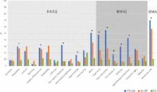
          Neurobehaviour was assessed at term-equivalent age in 149 PT<30, 200 MLPT and 200 term-born infants using the Neonatal Intensive Care UnitNetwork Neurobehavioral Scale (NNNS), the Hammersmith Neonatal Neurological Examination (HNNE) and Prechtl’s Qualitative Assessment of General Movements (GMA). A subset of 110 PT<30 and 198 MLPT infants had concurrent brain MRI.

          Results

          Proportions with abnormal neurobehaviour on the NNNS and the HNNE, and abnormal GMA all increased with decreasing gestational age. Higher brain MRI abnormality scores in some regions were associated with suboptimal neurobehaviour on the NNNS and HNNE. The relationships between brain MRI abnormality scores and suboptimal neurobehaviour were similar in both PT<30 and MLPT infants. The relationship between brain MRI abnormality scores and abnormal GMA was stronger in PT<30 infants.

          Conclusions

          There was a continuum of neurobehaviour across gestational ages. The relationships between brain abnormality scores and suboptimal neurobehaviour provide evidence that neurobehavioural assessments offer insight into the integrity of the developing brain, and may be useful in earlier identification of the highest-risk infants.

          Related collections

          Most cited references33

          • Record: found
          • Abstract: found
          • Article: not found

          New MR imaging assessment tool to define brain abnormalities in very preterm infants at term.

          WM injury is the dominant form of injury in preterm infants. However, other cerebral structures, including the deep gray matter and the cerebellum, can also be affected by injury and/or impaired growth. Current MR imaging injury assessment scales are subjective and are challenging to apply. Thus, we developed a new assessment tool and applied it to MR imaging studies obtained from very preterm infants at term age. MR imaging scans from 97 very preterm infants (< 30 weeks' gestation) and 22 healthy term-born infants were evaluated retrospectively. The severity of brain injury (defined by signal abnormalities) and impaired brain growth (defined with biometrics) was scored in the WM, cortical gray matter, deep gray matter, and cerebellum. Perinatal variables for clinical risks were collected. In very preterm infants, brain injury was observed in the WM (n=23), deep GM (n=5), and cerebellum (n=23). Combining measures of injury and impaired growth showed moderate to severe abnormalities most commonly in the WM (n=38) and cerebellum (n=32) but still notable in the cortical gray matter (n=16) and deep gray matter (n=11). WM signal abnormalities were associated with a reduced deep gray matter area but not with cerebellar abnormality. Intraventricular and/or parenchymal hemorrhage was associated with cerebellar signal abnormality and volume reduction. Multiple clinical risk factors, including prolonged intubation, prolonged parenteral nutrition, postnatal corticosteroid use, and postnatal sepsis, were associated with increased global abnormality on MR imaging. Very preterm infants demonstrate a high prevalence of injury and growth impairment in both the WM and gray matter. This MR imaging scoring system provides a more comprehensive and objective classification of the nature and extent of abnormalities than existing measures.
            Bookmark
            • Record: found
            • Abstract: found
            • Article: not found

            Neonatal intensive care unit stress is associated with brain development in preterm infants.

            Although many perinatal factors have been linked to adverse neurodevelopmental outcomes in very premature infants, much of the variation in outcome remains unexplained. The impact on brain development of 1 potential factor, exposure to stressors in the neonatal intensive care unit, has not yet been studied in a systematic, prospective manner. In this prospective cohort study of infants born at <30 weeks gestation, nurses were trained in recording procedures and cares. These recordings were used to derive Neonatal Infant Stressor Scale scores, which were employed to measure exposure to stressors. Magnetic resonance imaging (brain metrics, diffusion, and functional magnetic resonance imaging) and neurobehavioral examinations at term equivalent postmenstrual age were used to assess cerebral structure and function. Simple and partial correlations corrected for confounders, including immaturity and severity of illness, were used to explore these relations. Exposure to stressors was highly variable, both between infants and throughout a single infant's hospital course. Exposure to a greater number of stressors was associated with decreased frontal and parietal brain width, altered diffusion measures and functional connectivity in the temporal lobes, and abnormalities in motor behavior on neurobehavioral examination. Exposure to stressors in the Neonatal Intensive Care Unit is associated with regional alterations in brain structure and function. Further research into interventions that may decrease or mitigate exposure to stressors in the neonatal intensive care unit is warranted. Copyright © 2011 American Neurological Association.
              Bookmark
              • Record: found
              • Abstract: found
              • Article: not found

              Brain injury and altered brain growth in preterm infants: predictors and prognosis.

              To define the nature and frequency of brain injury and brain growth impairment in very preterm (VPT) infants by using MRI at term-equivalent age and to relate these findings to perinatal risk factors and 2-year neurodevelopmental outcomes. MRI scans at term-equivalent age from 3 VPT cohorts (n = 325) were reviewed. The severity of brain injury, including periventricular leukomalacia and intraventricular and cerebellar hemorrhage, was graded. Brain growth was assessed by using measures of biparietal width (BPW) and interhemispheric distance. Neurodevelopmental outcome at age 2 years was assessed across all cohorts (n = 297) by using the Bayley Scales of Infant Development, Second Edition (BSID-II) or Bayley Scales of Infant and Toddler Development, Third Edition (Bayley-III), and evaluation for cerebral palsy. Of 325 infants, 107 (33%) had some grade of brain injury and 33 (10%) had severe injury. Severe brain injury was more common in infants with lower Apgar scores, necrotizing enterocolitis, inotropic support, and patent ductus arteriosus. Severe brain injury was associated with delayed cognitive and motor development and cerebral palsy. Decreased BPW was related to lower gestational age, inotropic support, patent ductus arteriosus, necrotizing enterocolitis, prolonged parenteral nutrition, and oxygen at 36 weeks and was associated with delayed cognitive development. In contrast, increased interhemispheric distance was related to male gender, dexamethasone use, and severe brain injury. It was also associated with reduced cognitive development, independent of BPW. At term-equivalent age, VPT infants showed both brain injury and impaired brain growth on MRI. Severe brain injury and impaired brain growth patterns were independently associated with perinatal risk factors and delayed cognitive development. Copyright © 2014 by the American Academy of Pediatrics.
                Bookmark

                Author and article information

                Journal
                BMJ Paediatr Open
                BMJ Paediatr Open
                bmjpo
                bmjpo
                BMJ Paediatrics Open
                BMJ Publishing Group (BMA House, Tavistock Square, London, WC1H 9JR )
                2399-9772
                2017
                5 October 2017
                : 1
                : 1
                : e000136
                Affiliations
                [1 ]Murdoch Children’s Research Institute , Melbourne, Victoria, Australia
                [2 ]departmentNewborn Research , The Royal Women’s Hospital , Melbourne, Victoria, Australia
                [3 ]departmentDepartment of Paediatrics , Monash University , Melbourne, Victoria, Australia
                [4 ]Paediatric Infant Perinatal Emergency Retrieval (PIPER), The Royal Children’s Hospital , Melbourne, Victoria, Australia
                [5 ]departmentDepartment of Obstetrics and Gynaecology , University of Melbourne , Melbourne, Victoria, Australia
                [6 ]departmentDepartment of Paediatrics , University of Melbourne , Melbourne, Victoria, Australia
                [7 ]Florey Institute of Neuroscience and Mental Health , Melbourne, Victoria, Australia
                [8 ]Monash Institute of Cognitive and Clinical Neurosciences, Monash University , Melbourne, Victoria, Australia
                [9 ]departmentDepartment of Physiotherapy , University of Melbourne , Melbourne, Victoria, Australia
                Author notes
                [Correspondence to ] and Dr Alicia J Spittle; aspittle@ 123456unimelb.edu.au
                Article
                bmjpo-2017-000136
                10.1136/bmjpo-2017-000136
                5862173
                2f758f1e-bbb5-4eff-b05e-27bcd99df42b
                © Article author(s) (or their employer(s) unless otherwise stated in the text of the article) 2017. All rights reserved. No commercial use is permitted unless otherwise expressly granted.

                This is an Open Access article distributed in accordance with the Creative Commons Attribution Non Commercial (CC BY-NC 4.0) license, which permits others to distribute, remix, adapt, build upon this work non-commercially, and license their derivative works on different terms, provided the original work is properly cited and the use is non-commercial. See: http://creativecommons.org/licenses/by-nc/4.0/

                History
                : 31 May 2017
                : 06 September 2017
                : 12 September 2017
                Categories
                Original Article
                1506
                Custom metadata
                unlocked

                neurodevelopment
                neurodevelopment

                Comments

                Comment on this article