8
views
0
recommends
+1 Recommend
0 collections
    0
    shares
      • Record: found
      • Abstract: found
      • Article: found
      Is Open Access

      Epstein Barr Virus Associated Lymphomas and Epithelia Cancers in Humans

      review-article

      Read this article at

      Bookmark
          There is no author summary for this article yet. Authors can add summaries to their articles on ScienceOpen to make them more accessible to a non-specialist audience.

          Abstract

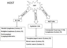
          Epstein Barr virus (EBV) is a cosmopolitan oncogenic virus, infecting about 90% of the world's population and it is associated to tumors originating from both epithelia and hematopoietic cells. Transmission of the virus is mainly through oral secretions; however, transmission through organ transplantation and blood transfusion has been reported. In order to evade immune recognition, EBV establishes latent infection in B lymphocytes where it expresses limited sets of proteins called EBV transcription programs (ETPs), including six nuclear antigens (EBNAs), three latent membrane proteins (LMP), and untranslated RNA called EBV encoded RNA (EBER), shown to efficiently transform B cells into lymphoblastic cells. These programs undergo different patterns of expression which determine the occurrence of distinct types of latency in the pathogenesis of a particular tumor. Hematopoietic cell derived tumors include but not limited to Burkitt's lymphoma, Hodgkin lymphoma, post-transplant lymphoproliferative disorders, and natural killer (NK)/T cell lymphoma. EBV undergoes lytic infection in epithelia cells for amplification of the viral particle for transmission where it expresses lytic stage genes. However, for reasons yet to be unveiled, EBV switches from the expression of lytic stage genes to the expression of ETPs in epithelia cells. The expression of the ETPs lead to the transformation of epithelia cells into permanently proliferating cells, resulting in epithelia cell derived malignancies such as nasopharyngeal cancer, gastric cancer, and breast cancer. In this review, we have summarized the current updates on EBV associated epithelial and B cell-derived malignancies, and the role of EBV latency gene products in the pathogenesis of the cancers, and have suggested areas for future studies when considering therapeutic measures

          Related collections

          Most cited references146

          • Record: found
          • Abstract: found
          • Article: not found

          Activation of NK cells and T cells by NKG2D, a receptor for stress-inducible MICA.

          Stress-inducible MICA, a distant homolog of major histocompatibility complex (MHC) class I, functions as an antigen for gammadelta T cells and is frequently expressed in epithelial tumors. A receptor for MICA was detected on most gammadelta T cells, CD8+ alphabeta T cells, and natural killer (NK) cells and was identified as NKG2D. Effector cells from all these subsets could be stimulated by ligation of NKG2D. Engagement of NKG2D activated cytolytic responses of gammadelta T cells and NK cells against transfectants and epithelial tumor cells expressing MICA. These results define an activating immunoreceptor-MHC ligand interaction that may promote antitumor NK and T cell responses.
            Bookmark
            • Record: found
            • Abstract: found
            • Article: not found

            A biologic definition of Burkitt's lymphoma from transcriptional and genomic profiling.

            The distinction between Burkitt's lymphoma and diffuse large-B-cell lymphoma is unclear. We used transcriptional and genomic profiling to define Burkitt's lymphoma more precisely and to distinguish subgroups in other types of mature aggressive B-cell lymphomas. We performed gene-expression profiling using Affymetrix U133A GeneChips with RNA from 220 mature aggressive B-cell lymphomas, including a core group of 8 Burkitt's lymphomas that met all World Health Organization (WHO) criteria. A molecular signature for Burkitt's lymphoma was generated, and chromosomal abnormalities were detected with interphase fluorescence in situ hybridization and array-based comparative genomic hybridization. We used the molecular signature for Burkitt's lymphoma to identify 44 cases: 11 had the morphologic features of diffuse large-B-cell lymphomas, 4 were unclassifiable mature aggressive B-cell lymphomas, and 29 had a classic or atypical Burkitt's morphologic appearance. Also, five did not have a detectable IG-myc Burkitt's translocation, whereas the others contained an IG-myc fusion, mostly in simple karyotypes. Of the 176 lymphomas without the molecular signature for Burkitt's lymphoma, 155 were diffuse large-B-cell lymphomas. Of these 155 cases, 21 percent had a chromosomal breakpoint at the myc locus associated with complex chromosomal changes and an unfavorable clinical course. Our molecular definition of Burkitt's lymphoma clarifies and extends the spectrum of the WHO criteria for Burkitt's lymphoma. In mature aggressive B-cell lymphomas without a gene signature for Burkitt's lymphoma, chromosomal breakpoints at the myc locus were associated with an adverse clinical outcome. Copyright 2006 Massachusetts Medical Society.
              Bookmark
              • Record: found
              • Abstract: found
              • Article: not found

              Clinical differences between nasal and extranasal natural killer/T-cell lymphoma: a study of 136 cases from the International Peripheral T-Cell Lymphoma Project.

              Among 1153 new adult cases of peripheral/T-cell lymphoma from 1990-2002 at 22 centers in 13 countries, 136 cases (11.8%) of extranodal natural killer (NK)/T-cell lymphoma were identified (nasal 68%, extranasal 26%, aggressive/unclassifiable 6%). The disease frequency was higher in Asian than in Western countries and in Continental Asia than in Japan. There were no differences in age, sex, ethnicity, or immunophenotypic profile between the nasal and extranasal cases, but the latter had more adverse clinical features. The median overall survival (OS) was better in nasal compared with the extranasal cases in early- (2.96 vs 0.36 years, P < .001) and late-stage disease (0.8 vs 0.28 years, P = .031). The addition of radiotherapy for early-stage nasal cases yielded survival benefit (P = .045). Among nasal cases, both the International Prognostic Index (P = .006) and Korean NK/T-cell Prognostic Index (P < .001) were prognostic. In addition, Ki67 proliferation greater than 50%, transformed tumor cells greater than 40%, elevated C-reactive protein level (CRP), anemia (< 11 g/dL) and thrombocytopenia (< 150 x 10(9)/L) predicts poorer OS for nasal disease. No histologic or clinical feature was predictive in extranasal disease. We conclude that the clinical features and treatment response of extranasal NK/T-cell lymphoma are different from of those of nasal lymphoma. However, the underlying features responsible for these differences remain to be defined.
                Bookmark

                Author and article information

                Journal
                J Cancer
                J Cancer
                jca
                Journal of Cancer
                Ivyspring International Publisher (Sydney )
                1837-9664
                2020
                17 January 2020
                : 11
                : 7
                : 1737-1750
                Affiliations
                [1 ]Department of Biochemistry, Cell and Molecular Biology, University of Ghana, Legon, Accra, Ghana
                [2 ]West African Center for Cell Biology of Infectious Pathogens (WACCBIP), University of Ghana, Legon, Accra, Ghana
                [3 ]Department of Biochemistry, University of Sussex, Brighton, U.K
                Author notes
                ✉ Corresponding author: Osbourne Quaye, Department of Biochemistry, Cell and Molecular Biology, University of Ghana, Volta Road, P. O. Box LG 54, Legon, Accra-Ghana; Tel: +233-2611-70904.

                Competing Interests: The authors have declared that no competing interest exists.

                Article
                jcav11p1737
                10.7150/jca.37282
                7052849
                32194785
                2fa5cad4-8c0b-4601-ba0e-f3eda880cce6
                © The author(s)

                This is an open access article distributed under the terms of the Creative Commons Attribution License ( https://creativecommons.org/licenses/by/4.0/). See http://ivyspring.com/terms for full terms and conditions.

                History
                : 3 June 2019
                : 1 December 2019
                Categories
                Review

                Oncology & Radiotherapy
                epstein barr virus,lymphomas,epithelial cancers,latency program
                Oncology & Radiotherapy
                epstein barr virus, lymphomas, epithelial cancers, latency program

                Comments

                Comment on this article