51
views
0
recommends
+1 Recommend
0 collections
    0
    shares
      • Record: found
      • Abstract: found
      • Article: found
      Is Open Access

      Anatomical Study of Intrahemispheric Association Fibers in the Brains of Capuchin Monkeys ( Sapajus sp.)

      research-article

      Read this article at

      Bookmark
          There is no author summary for this article yet. Authors can add summaries to their articles on ScienceOpen to make them more accessible to a non-specialist audience.

          Abstract

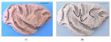
          Previous studies suggest that the complexity of fiber connections in the brain plays a key role in the evolutionary process of the primate brain and behaviors. The patterns of brain fiber systems have been studied in detail in many nonhuman primates, but not in Sapajus sp. Behavioral studies indicated that Sapajus sp. (bearded capuchins) show highly cognitive behaviors such as tool use comparable to those in other nonhuman primates. To compare the brain fiber systems in capuchins with those in other nonhuman primates and humans, the intrahemispheric fibers systems in 24 cerebral hemispheres of Sapajus were dissected by a freezing-thawing procedure. Dissection of the hemispheres in lateral view indicated short arcuate fibers, uncinate fasciculus, and inferior longitudinal fasciculus, while that in a medial view indicated short arcuate fibers, the cingulum united with the superior longitudinal fasciculus, and inferior longitudinal fasciculus. The results showed that the fiber systems in Sapajus are comparable to those in rhesus and humans, except for a lack of independent superior longitudinal fasciculus and cingulum in Sapajus.

          Related collections

          Most cited references43

          • Record: found
          • Abstract: found
          • Article: not found

          Limbic connections of the orbital and medial prefrontal cortex in macaque monkeys.

          Previous studies have shown that the orbital and medial prefrontal cortex (OMPFC) is extensively connected with medial temporal and cingulate limbic structures. In this study, the organization of these projections was defined in relation to architectonic areas within the OMPFC. All of the limbic structures were substantially connected with the following posterior and medial orbital areas: the posteromedial, medial, intermediate, and lateral agranular insular areas (Iapm, Iam, Iai, and Ial, respectively) and areas 11m, 13a, 13b, 14c and 14r. In contrast, lateral orbital areas 12o, 12m, and 12l and medial wall areas 24a,b and 32 were primarily connected with the amygdala, the temporal pole, and the cingulate cortex. Data were not obtained on the posteroventral medial wall. Three distinct projections were recognized from the basal amygdaloid nucleus: 1) The dorsal part projected to area 12l; 2) the ventromedial part projected to most areas in the posterior and medial orbital cortex except for area Iai, 12o, 13a, and 14c; and 3) the ventrolateral part projected to orbital areas 12o, Iai, 13a, 14c, and to the medial wall areas. The accessory basal and lateral amygdaloid nuclei projected most strongly to areas in the posterior and medial orbital cortex. The medial, anterior cortical, and central amygdaloid nuclei and the periamygdaloid cortex were connected with the posterior orbital areas. The projection from the hippocampus originated from the rostral subiculum and terminated in the medial orbital areas. The same region was reciprocally connected with the anteromedial nucleus of the thalamus, which received input from the rostral subiculum. The parahippocampal cortical areas (including the temporal polar, entorhinal, perirhinal, and posterior parahippocampal cortices) were primarily connected with posterior and medial orbital areas, with some projections to the dorsal part of the medial wall. The rostral cingulate cortex sent fibers to the medial wall, to the medial orbital areas, and to lateral areas 12o, 12r, and Iai. The posterior cingulate gyrus, including the caudomedial lobule, was especially strongly connected with area 11m.
            Bookmark
            • Record: found
            • Abstract: found
            • Article: not found

            Architecture and intrinsic connections of the prefrontal cortex in the rhesus monkey.

            An investigation of the architectonic organization and intrinsic connections of the prefrontal cortex was conducted in rhesus monkeys. Cytoarchitectonic analysis indicates that in the prefrontal cortex there are two trends of gradual change in laminar characteristics that can be traced from limbic periallocortex towards isocortical areas. The stepwise change in laminar features is characterized by the emergence and gradual increase in the width of granular layer IV, by an increase in the size of pyramidal cells in layers III and V, and by a higher cell-packing density in the supragranular layers. Myeloarchitectonic analysis reveals that the limbic areas are poorly myelinated, adjacent areas have a diffuse myelin content confined to the deep layers, and in isocortices the myelinated fibers are distributed in organized horizontal bands (of Bail-larger) and a vertical plexus. Using the above architectonic criteria, we observed that one of the architectonic trends takes a radial basoventral course from the periallocortex in the caudal orbitofrontal region to the adjacent proisocortex and then to area 13. The next stage of architectonic regions includes orbital areas 12, 11, and 14, which is followed by area 10, lateral area 12, and the rostral part of ventral area 46. The last group includes the caudal part of ventral area 46 and ventral area 8. The other trend takes a mediodorsal course from the periallocortex around the rostral portion of the corpus callosum to the adjacent proisocortical areas 24, 25, and 32 and then to the medially situated isocortical areas 9, 10, and 14. The next stage includes lateral areas 10 and 9 and the rostral part of dorsal area 46. The last group includes the caudal part of dorsal area 46 and dorsal area 8. The interconnections of subdivisions of the basoventral and mediodorsal cortices were studied with the aid of anterograde and retrograde tracers. Within each trend a given area projects in two directions: to adjoining regions belonging to succeeding architectonic stages on the one hand, and to nearby regions from the preceding architectonic stage on the other. In each direction there is more than one region involved in this projection system, paralleling the radial nature of architectonic change. Periallo- and proisocortices have widespread intrinsic connections, whereas isocortices situated at a distance from limbic areas, such as area 8, have restricted connections. Most interconnections are limited to areas within the same architectonic trend. However, there are links between cortices from the two trends, and these seem to occur between areas that are at a similar stage of architectonic differentiation.(ABSTRACT TRUNCATED AT 400 WORDS)
              Bookmark
              • Record: found
              • Abstract: found
              • Article: not found

              Beyond the arcuate fasciculus: consensus and controversy in the connectional anatomy of language.

              The growing consensus that language is distributed into large-scale cortical and subcortical networks has brought with it an increasing focus on the connectional anatomy of language, or how particular fibre pathways connect regions within the language network. Understanding connectivity of the language network could provide critical insights into function, but recent investigations using a variety of methodologies in both humans and non-human primates have provided conflicting accounts of pathways central to language. Some of the pathways classically considered language pathways, such as the arcuate fasciculus, are now argued to be domain-general rather than specialized, which represents a radical shift in perspective. Other pathways described in the non-human primate remain to be verified in humans. In this review, we examine the consensus and controversy in the study of fibre pathway connectivity for language. We focus on seven fibre pathways-the superior longitudinal fasciculus and arcuate fasciculus, the uncinate fasciculus, extreme capsule, middle longitudinal fasciculus, inferior longitudinal fasciculus and inferior fronto-occipital fasciculus-that have been proposed to support language in the human. We examine the methods in humans and non-human primate used to investigate the connectivity of these pathways, the historical context leading to the most current understanding of their anatomy, and the functional and clinical correlates of each pathway with reference to language. We conclude with a challenge for researchers and clinicians to establish a coherent framework within which fibre pathway connectivity can be systematically incorporated to the study of language.
                Bookmark

                Author and article information

                Journal
                Biomed Res Int
                Biomed Res Int
                BMRI
                BioMed Research International
                Hindawi Publishing Corporation
                2314-6133
                2314-6141
                2015
                29 November 2015
                : 2015
                : 648128
                Affiliations
                1Department of Biology, Academic Areas, Federal Institute of Goiás, 75131-45 Anápolis, GO, Brazil
                2System Emotional Science, Graduate School of Medicine and Pharmaceutical Sciences, University of Toyama, Toyama 930-0194, Japan
                3Laboratory of Anthropology, Biochemistry, Neuroscience and Primate Behavior (LABINECOP), Federal University of Tocantins, 77001-090 Palmas, TO, Brazil
                4School of Medicine, University of Brasília, 70910-900 Brasília, DF, Brazil
                5Behavioral Neurology Unit, Hospital das Clínicas, Federal University of Goiás, 74605-020 Goiânia, GO, Brazil
                Author notes
                *Kellen Christina Malheiros Borges: kellen.borges@ 123456ifg.edu.br

                Academic Editor: Gerald J. Wyckoff

                Article
                10.1155/2015/648128
                4676999
                26693488
                311c1388-1da5-4e61-a896-35d22e52e486
                Copyright © 2015 Kellen Christina Malheiros Borges et al.

                This is an open access article distributed under the Creative Commons Attribution License, which permits unrestricted use, distribution, and reproduction in any medium, provided the original work is properly cited.

                History
                : 8 September 2015
                : 6 November 2015
                : 15 November 2015
                Categories
                Research Article

                Comments

                Comment on this article