2
views
0
recommends
+1 Recommend
0 collections
    0
    shares
      • Record: found
      • Abstract: found
      • Article: found
      Is Open Access

      Liver abscess secondary to fishbone ingestion: case report and review of the literature

      case-report
      , ,
      Journal of Surgical Case Reports
      Oxford University Press

      Read this article at

      Bookmark
          There is no author summary for this article yet. Authors can add summaries to their articles on ScienceOpen to make them more accessible to a non-specialist audience.

          Abstract

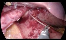
          We report a rare silent migration of a fishbone into the liver and review the relevant literature. A 56-year-old man presented with a 2-day history of dull epigastric pain and raised inflammatory markers. Computerized tomography scan revealed a 4-cm abscess in the left lobe of the liver, with a linear radio-dense foreign body within the collection. At laparoscopy the hepatogastric fistula was disconnected. The fishbone was retrieved from the liver. Gastrostomy was closed with an omental patch. The patient had an uneventful recovery. Fifty-two cases of liver abscess secondary to enterohepatic fishbone migration were reported with over two-thirds presenting with a left-lobe abscess. There was marked variability in the management of liver abscess in the setting of fishbone migration-summarized in table. We believe that laparoscopic drainage of the abscess and extraction of the foreign body offer control of the source of sepsis and diminishes recurrence, whilst having a low-risk profile.

          Related collections

          Most cited references49

          • Record: found
          • Abstract: found
          • Article: not found

          Hepatic abscess induced by foreign body: case report and literature review.

          Hepatic abscess due to perforation of the gastrointestinal tract caused by ingested foreign bodies is uncommon. Pre-operative diagnosis is difficult as patients are often unaware of the foreign body ingestion and symptoms and imagiology are usually non-specific. The authors report a case of 62-year-old woman who was admitted with fever and abdominal pain. Further investigation revealed hepatic abscess, without resolution despite antibiotic therapy. A liver abscess resulting from perforation and intra-hepatic migration of a bone coming from the pilorum was diagnosed by surgery. The literature concerning foreign body-induced perforation of the gastrointestinal tract complicated by liver abscess is reviewed.
            Bookmark
            • Record: found
            • Abstract: found
            • Article: not found

            Gastrointestinal foreign bodies.

            Over a 20 year period 26 patients were admitted because of ingesting foreign bodies. Ten patients, mostly children, remembered swallowing a metallic foreign body. Most of the patients were asymptomatic and were admitted for observation. Sixteen patients underwent operation. The distal large bowel was the area most commonly involved, not the ileocecal region as in the reviews published a number of years ago. Blunt as well as pointed foreign bodies caused bowel perforations, probably through slow pressure necrosis. Most of the patients had signs of localized peritonitis.. Unlike previous studies, roentgenographic studies were found helpful in the preoperative diagnosis.
              Bookmark
              • Record: found
              • Abstract: found
              • Article: found
              Is Open Access

              Liver abscess caused by fish bone perforation of stomach wall treated by laparoscopic surgery: a case report

              Background Formation of a liver abscess due to gastrointestinal perforation by a foreign body is rare. In addition, there are few case reports on laparoscopic surgical treatment of a liver abscess caused by perforation of the gastrointestinal tract by a foreign body. Case presentation A 51-year-old man visited our hospital because of fever and anorexia. There were no physical findings except for fever. He had no comorbidities or surgical history. Laboratory tests showed increased inflammatory marker and liver enzyme levels. Abdominal ultrasonography showed a hypoechoic lesion in the left lobe of the liver. Abdominal contrast-enhanced computed tomography revealed an air-containing abscess in the left side of the liver and a high-density linear object. We diagnosed a liver abscess secondary to stomach perforation by a foreign body. Emergency laparoscopic surgery identified a fish bone in the abscess that formed between the stomach and liver. We succeeded in removing the fish bone laparoscopically. The patient was discharged without any postoperative complications on day 11. Conclusions A liver abscess secondary to perforation of the gastrointestinal tract by a foreign body usually requires surgical treatment. Foreign body removal is important to prevent recurrence of liver abscess. In cases with the foreign body located at the liver margin, a laparoscopic approach to the abscess is very useful.
                Bookmark

                Author and article information

                Journal
                J Surg Case Rep
                J Surg Case Rep
                jscr
                Journal of Surgical Case Reports
                Oxford University Press
                2042-8812
                February 2022
                15 February 2022
                15 February 2022
                : 2022
                : 2
                : rjac026
                Affiliations
                [1] Institute of Liver Studies , Kings College Hospital, London, UK
                Author notes
                Correspondence address. King’s College Hospital, Denmark Hill, London SE5 9RS, UK. Tel: +44-7740-705060; Email: Ameet.1.patel@ 123456kcl.ac.uk
                Article
                rjac026
                10.1093/jscr/rjac026
                8846943
                35178243
                31a429fb-4875-425e-aa83-8759ec734eac
                Published by Oxford University Press and JSCR Publishing Ltd. © The Author(s) 2022.

                This is an Open Access article distributed under the terms of the Creative Commons Attribution License ( https://creativecommons.org/licenses/by/4.0/), which permits unrestricted reuse, distribution, and reproduction in any medium, provided the original work is properly cited.

                History
                : 2 December 2021
                : 15 January 2022
                Page count
                Pages: 5
                Funding
                Funded by: Kings College London, DOI 10.13039/501100000764;
                Categories
                AcademicSubjects/MED00910
                jscrep/0
                Case Report

                Comments

                Comment on this article