174
views
0
recommends
+1 Recommend
0 collections
    0
    shares
      • Record: found
      • Abstract: found
      • Article: not found

      Crystal structure of the FimD usher bound to its cognate FimC:FimH substrate

      research-article

      Read this article at

      ScienceOpenPublisherPMC
      Bookmark
          There is no author summary for this article yet. Authors can add summaries to their articles on ScienceOpen to make them more accessible to a non-specialist audience.

          Abstract

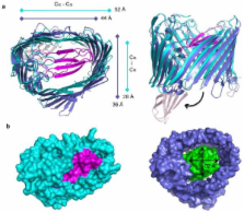
          Type 1 pili are the archetypal representative of a widespread class of adhesive multisubunit fibres in Gram-negative bacteria. During pilus assembly, subunits dock as chaperone-bound complexes to an usher, which catalyzes their polymerization and mediates pilus translocation across the outer membrane. We report the crystal structure of the full-length FimD usher bound to the FimC:FimH chaperone:adhesin complex and that of the unbound form of the FimD translocation domain. The FimD:FimC:FimH structure shows FimH inserted inside the FimD 24-stranded β-barrel translocation channel. FimC:FimH is held in place through interactions with the two C-terminal periplasmic domains of FimD, a binding mode confirmed in solution by electron paramagnetic resonance spectroscopy. To accommodate FimH, the usher plug domain is displaced from the barrel lumen to the periplasm, concomitant with a dramatic conformational change in the β-barrel. The N-terminal domain of FimD is observed in an ideal position to catalyse incorporation of a newly recruited chaperone:subunit complex. The FimD:FimC:FimH structure provides unique insights into the pilus subunit incorporation cycle, and captures the first view of a protein transporter in the act of secreting its cognate substrate.

          Related collections

          Most cited references34

          • Record: found
          • Abstract: found
          • Article: not found

          Shape complementarity at protein/protein interfaces.

          A new statistic Sc, which has a number of advantages over other measures of packing, is used to examine the shape complementarity of protein/protein interfaces selected from the Brookhaven Protein Data Bank. It is shown using Sc that antibody/antigen interfaces as a whole exhibit poorer shape complementarity than is observed in other systems involving protein/protein interactions. This result can be understood in terms of the fundamentally different evolutionary history of particular antibody/antigen associations compared to other systems considered, and in terms of the differing chemical natures of the interfaces.
            Bookmark
            • Record: found
            • Abstract: found
            • Article: not found

            The integration of macromolecular diffraction data.

            The objective of any modern data-processing program is to produce from a set of diffraction images a set of indices (hkls) with their associated intensities (and estimates of their uncertainties), together with an accurate estimate of the crystal unit-cell parameters. This procedure should not only be reliable, but should involve an absolute minimum of user intervention. The process can be conveniently divided into three stages. The first (autoindexing) determines the unit-cell parameters and the orientation of the crystal. The unit-cell parameters may indicate the likely Laue group of the crystal. The second step is to refine the initial estimate of the unit-cell parameters and also the crystal mosaicity using a procedure known as post-refinement. The third step is to integrate the images, which consists of predicting the positions of the Bragg reflections on each image and obtaining an estimate of the intensity of each reflection and its uncertainty. This is carried out while simultaneously refining various detector and crystal parameters. Basic features of the algorithms employed for each of these three separate steps are described, principally with reference to the program MOSFLM.
              Bookmark
              • Record: found
              • Abstract: found
              • Article: not found

              Dead-time free measurement of dipole-dipole interactions between electron spins.

              A four-pulse version of the pulse double electron-electron resonance (DEER) experiment is presented, which is designed for the determination of interradical distances on a nanoscopic length-scale. With the new pulse sequence electron-electron couplings can be studied without dead-time artifacts, so that even broad distributions of electron-electron distances can be characterized. A version of the experiment that uses a pulse train in the detection period exhibits improved signal-to-noise ratio. Tests on two nitroxide biradicals with known length indicate that the accessible range of distances extends from about 1.5 to 8 nm. The four-pulse DEER spectra of an ionic spin probe in an ionomer exhibit features due to probe molecules situated both on the same and on different ion clusters. The former feature provides information on the cluster size and is inaccessible with previous methods. Copyright 2000 Academic Press.
                Bookmark

                Author and article information

                Journal
                0410462
                6011
                Nature
                Nature
                Nature
                0028-0836
                1476-4687
                21 July 2011
                2 June 2011
                02 December 2011
                : 474
                : 7349
                : 49-53
                Affiliations
                [1 ]Institute of Structural and Molecular Biology, University College London and Birkbeck College, Malet Street, London, WC1E 7HX, UK
                [2 ]Structural & Molecular Microbiology, VIB - Vrije Universiteit Brussels, 1050 Brussels, Belgium
                [3 ]Biology Department, Brookhaven National Laboratory, Upton, NY 11973, USA
                [4 ]Department of Chemistry, University of York, York, YO10 5YW, UK
                [5 ]Center for Infectious Diseases and Department of Molecular Genetics & Microbiology, Stony Brook University, Stony Brook, NY 11794, USA
                [6 ]Department of Molecular Microbiology and Center for Women’s Infectious Disease Research, Washington University School of Medicine, Saint Louis, MO63110, USA
                [7 ]Department of Biochemistry and Cell Biology, Stony Brook University, Stony Brook, NY 11794, USA
                [8 ]London Centre for Nanotechnology, University College London, London WC1H 0AH, UK
                [9 ]Research Department of Structural and Molecular Biology, University College London, Gower Street, WC1E 6BT, UK
                Author notes
                Authors for correspondence and requests for materials. Gabriel Waksman at g.waksman@ 123456ucl.ac.uk or g.waksman@ 123456bbk.ac.uk , David Thanassi at david.thanassi@ 123456stonybrook.edu
                [*]

                These authors contributed equally to the work

                Author contribution statement. G.P. produced the FimD:FimC:FimH complex, grew the crystals of this complex, collected X-ray crystallographic data, and initiated the determination of the structure by Molecular Replacement, and participated in the building and refinement of the structure. H.R. produced the FimD:FimC:FimH complex, trained G.P., supervised the work, analysed the structures and wrote the paper. T.W. grew crystals of the FimD translocation domain, collected X-ray crystallographic data, and determined the structure. W.J.A. set up the DSE assay and prepared the samples for EPR. K.F.P. carried out the EPR experiments, which were analysed by K.F.P., M.B.A.K. and C.W.M.K. A.L. completed the structure determination of the FimD:FimC:FimH complex, built and refined the structure. N.S.H., E.V., J.S.P and B.F. cloned and purified the translocation domain of FimD, and cloned and analyzed the FimD CTD mutants. S.G. participated in the building and refinement of the FimD:FimC:FimH structure and analysed the structure. J.Y. carried out the native mass spectrometry experiments on the FimD:FimC:FimH complex. C.W.M.K supervised the EPR work. H.L., S.J.H. and D.G.T. supervised the work on apo-FimD, analysed the structures, and wrote the paper. G.W. supervised the work on FimD:FimC:FimH, analysed the structures, and wrote the paper.

                Article
                UKMS36012
                10.1038/nature10109
                3162478
                21637253
                3285e39a-f064-4bb6-8bd7-085b571b50a3

                Users may view, print, copy, download and text and data- mine the content in such documents, for the purposes of academic research, subject always to the full Conditions of use: http://www.nature.com/authors/editorial_policies/license.html#terms

                History
                Funding
                Funded by: Medical Research Council :
                Award ID: G0100442(58149) || MRC_
                Categories
                Article

                Uncategorized
                Uncategorized

                Comments

                Comment on this article