27
views
0
recommends
+1 Recommend
0 collections
    0
    shares
      • Record: found
      • Abstract: found
      • Article: found
      Is Open Access

      Infectious sacroiliitis: a retrospective, multicentre study of 39 adults

      research-article

      Read this article at

      Bookmark
          There is no author summary for this article yet. Authors can add summaries to their articles on ScienceOpen to make them more accessible to a non-specialist audience.

          Abstract

          Background

          Non-brucellar and non-tuberculous infectious sacroiliitis (ISI) is a rare disease, with misleading clinical signs that delay diagnosis. Most observations are based on isolated case reports or small case series. Our aim was to describe the clinical, bacteriological, and radiological characteristics of ISI, as well as the evolution of these arthritis cases under treatment.

          Methods

          This retrospective study included all ISI cases diagnosed between 1995 and 2011 in eight French rheumatology departments. ISI was diagnosed if sacroiliitis was confirmed bacteriologically or, in the absence of pathogenic agents, if clinical, biological, and radiological data was compatible with this diagnosis and evolution was favourable under antibiotic therapy.

          Results

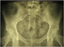
          Overall, 39 cases of ISI were identified in adults, comprising 23 women and 16 men, with a mean age at diagnosis of 39.7 ± 18.1 years. The left sacroiliac joint (SI) was affected in 59% of cases, with five cases occurring during the post-partum period. Lumbogluteal pain was the most common symptom (36/39). Manipulations of the SI joint were performed in seven patients and were always painful. Mean score for pain using the visual analogue score was 7.3/10 at admission, while 16 patients were febrile at diagnosis. No risk factor was found for 30.7% of patients. A diagnosis of ISI was only suspected in five cases at admission. The mean time to diagnosis was long, being 43.3 ± 69.1 days on average. Mean C-reactive protein was 149.7 ± 115.3 mg/l, and leukocytosis (leukocytes ≥ 10 G/l) was uncommon (n = 15) (mean level of leukocytes 10.4 ± 3.5 G/l). Radiographs (n = 33) were abnormal in 20 cases, revealing lesions of SI, while an abdominopelvic computed tomography (CT) scan (n = 27) was abnormal in 21 cases, suggesting arthritis of the SI joints in 13 cases (48.1%) and a psoas abscess in eight. Bone scans (n = 14) showed hyperfixation of the SI in 13 cases. Magnetic resonance imaging (MRI) (n = 27), when focused on the SI (n = 25), directed towards the diagnosis to ISI in 25 cases. Pathogenic agents were isolated in 33 cases (84.6%) by means of articular puncture (n = 16), blood culture (n = 14), cytobacteriological examination of urine (n = 2), or puncture of the psoas (n = 1).

          Gram-positive cocci were the mostly isolated common bacteria, with a predominance of staphylococci (n = 21). The most frequently isolated gram-negative bacillus was Pseudomonas aeruginosa (n = 3). Evolution was favourable in 37 out of 39 patients under prolonged antibiotic therapy (mean duration 3.01 ± 1.21 months).

          Conclusion

          Our series confirmed that the clinical manifestations of ISI usually lead to delayed diagnosis. Based on our results, we suggest performing an MRI of the spine and SI in clinical situations characterised by lumbogluteal pain and symptoms of an infectious disease, such as fever.

          Related collections

          Most cited references24

          • Record: found
          • Abstract: found
          • Article: not found

          Pyogenic sacroiliitis--a comparison between paediatric and adult patients.

          Pyogenic sacroiliitis is a rare cause of hip pain and fever. We aim to report a series of 33 patients with pyogenic sacroiliitis and to investigate the differences among paediatric and adult cases. Clinical and laboratory data of 33 cases who were admitted to the emergency department with a confirmed discharge diagnosis of pyogenic sacroiliitis between 1996 and 2005 were assessed. All patients were divided into paediatric and adult groups by the age of 15 yr. The features of pyogenic sacroiliitis and clinical outcome were compared among the two groups. Several factors were analysed including gender, age, clinical signs and symptoms, laboratory tests, radiological examinations and scintigraphy. Among the all included patients, females were attacked more frequently than males (3: 1). One-third of patients had concurrent infections, of which urinary tract infections were the most common (41.6%). Compared with adult patients, paediatric patients tend to have fewer comorbid immunocompromized conditions, fewer concurrent infections, more equality in gender distribution and more presentations of weight bearing difficulty. Staphylococcus aureus was the main blood culture isolate from paediatric patients (80%), but only accounted for half of those from adult patients. Group B streptococcus and Salmonella spp. were not uncommon in the adult patients. Scintigraphic bone scan has the highest sensitivity (93.3%) and remains the image modality of choice. When local abscess formation is suspected, computed tomography or magnetic resonance imaging may be the preferred method used for examination. This case series should alert the physicians to the possibility of pyogenic sacroiliitis and the difference between paediatric and adult patients.
            Bookmark
            • Record: found
            • Abstract: found
            • Article: not found

            Arthritis in leprosy.

            Leprosy, a chronic granulomatous infection caused by Mycobacterium leprae, classically presents with cutaneous and neurological manifestations. Musculoskeletal involvement though third most common is underdiagnosed and underreported. It may manifest in the form of Charcot's arthropathy, acute symmetrical polyarthritis or swollen hands and feet syndrome during lepra reactions, insidious-onset chronic symmetrical polyarthritis mimicking RA or as isolated tenosynovitis or tenosynovitis associated with arthritis or neuropathy. At times, articular involvement may be the sole presenting manifestation even without cutaneous lesions. Other rheumatological manifestations occasionally reported are enthesitis, sacroiliitis, cryoglobulinaemic vasculitis and DM. With increasing travel of population between tropical and temperate zones, it is likely that rheumatology clinics in countries free of leprosy may come across cases of leprosy with rheumatological manifestations. Delay in diagnosis and management may be detrimental and may result in deformities and loss of function. Not only this, but recent reports of leprosy being diagnosed in native white populations following anti-TNF-α therapy should alert rheumatologists across the globe to be more familiar with this disease. This review is aimed at presenting a comprehensive clinical scenario of various rheumatological manifestations of leprosy to sensitize rheumatologists and physicians across the continents.
              Bookmark
              • Record: found
              • Abstract: found
              • Article: not found

              Pyogenic infection of the sacroiliac joint. Case reports and review of the literature.

              Three cases of pyogenic sacroiliitis are described, and the English literature from 1878 to 1990 reviewed, for a total of 166 cases. In 1 patient the source of infection was identified at the site of an intravenous line; 1 patient had 2 risk factors for developing the disease (pregnancy and intravenous drug use); and a third patient had no source of infection and no associated risk factors. The diagnosis of pyogenic sacroiliitis was made in each patient by history, physical examination, and positive skeletal scintigraphy or computed tomography of the sacroiliac joint. The infectious agent causing septic arthritis was identified by fine-needle aspiration of the sacroiliac joint under fluoroscopic guidance. Two of the 3 patients also had an open biopsy of the sacroiliac joint--one to confirm the organism causing septic arthritis, and the other for surgical drainage of the infected sacroiliac joint. Cultures from all 3 patients grew organisms uncommon for this disease, and all were treated for 6 weeks with intravenous antibiotics. In all patients pain diminished after treatment. Pyogenic sacroiliitis is a relatively rare condition (1-2 cases reported/year) that may be clinically difficult to diagnose unless the clinician is familiar with the disease. A prompt diagnosis can prevent significant morbidity and reduce serious complication. Major predisposing factors include intravenous drug use, trauma, or an identifiable focus of infection elsewhere, but 44% of patients have no predisposing or associated factors identified. Most patients present with an acute febrile illness with pain in the buttocks and pain on movement that stresses the affected sacroiliac joint. There is no specific blood test which points to the diagnosis of pyogenic sacroiliitis, although the erythrocyte sedimentation rate may be greater than 100 mm/hr. The diagnostic procedure of choice is bone scan with attention to the early perfusion phase, which usually localizes the affected sacroiliac joint. Unilateral involvement is the rule. In patients whose blood cultures fail to reveal a causative organism, fluoroscopic guided fine-needle aspiration of the sacroiliac joint under general anesthesia may help to identify the organism. If all cultures are negative, open biopsy of the sacroiliac joint may be required. Open biopsy should also be done if sequestration or an abscess is formed, or if the patient fails to respond to antibiotic therapy.(ABSTRACT TRUNCATED AT 400 WORDS)
                Bookmark

                Author and article information

                Journal
                BMC Infect Dis
                BMC Infect. Dis
                BMC Infectious Diseases
                BioMed Central
                1471-2334
                2012
                15 November 2012
                : 12
                : 305
                Affiliations
                [1 ]Service de Médecine Interne, Hôpital G. Montpied, CHU de Clermont-Ferrand, 58 rue Montalembert, Cedex 1, Clermont-Ferrand 63003, France
                [2 ]Service de Rhumatologie, Hôpital R. Salengro, CHU de Lille, Lille, France
                [3 ]Service de Rhumatologie, Hôpital G. Montpied, CHU de Clermont-Ferrand, Clermont-Ferrand, France
                [4 ]Service de Rhumatologie A, Université Paris Descartes, Hôpital Cochin, Paris, France
                [5 ]Service de Rhumatologie, Hôpital de la Croix St Simon, Paris, France
                [6 ]Service de Rhumatologie, Hôpital Sud, CHU de Grenoble, Grenoble, France
                [7 ]Service de Rhumatologie, Hôpital Nord, CHU de St Etienne, St Etienne, France
                [8 ]Service de Rhumatologie, Centre Hospitalier, Le Mans, France
                [9 ]Service de Rhumatologie, Hôpital J. Lacarin, CH de Vichy, Vichy, France
                Article
                1471-2334-12-305
                10.1186/1471-2334-12-305
                3519695
                23153120
                32ef719e-e27e-438f-b790-38b3d92da32f
                Copyright ©2012 Hermet et al.; licensee BioMed Central Ltd.

                This is an Open Access article distributed under the terms of the Creative Commons Attribution License ( http://creativecommons.org/licenses/by/2.0), which permits unrestricted use, distribution, and reproduction in any medium, provided the original work is properly cited.

                History
                : 23 April 2012
                : 8 November 2012
                Categories
                Research Article

                Infectious disease & Microbiology
                Infectious disease & Microbiology

                Comments

                Comment on this article