40
views
0
recommends
+1 Recommend
0 collections
    0
    shares
      • Record: found
      • Abstract: found
      • Article: not found

      T 1–T 2 Dual-modal MRI contrast agents based on superparamagnetic iron oxide nanoparticles with surface attached gadolinium complexes

      research-article

      Read this article at

      ScienceOpenPublisherPMC
      Bookmark
          There is no author summary for this article yet. Authors can add summaries to their articles on ScienceOpen to make them more accessible to a non-specialist audience.

          Abstract

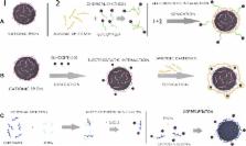
          Dual-mode MRI contrast agents consisting of superparamagnetic iron oxide nanoparticle (SPION) cores and gadolinium ions associated with the ionic chitosan protecting layer were synthesized and studied. Gadolinium ions were introduced into the coating layer via direct complex formation on the nanoparticles surface, covalent attachment or electrostatically driven deposition of the preformed Gd complex. The modified SPIONs having hydrodynamic diameters ca. 100 nm form stable, well-defined dispersions in water and have excellent magnetic properties. Physiochemical properties of those new materials were characterized using e.g., FTIR spectroscopy, dynamic light scattering, X-ray fluorescence, TEM, and vibrating sample magnetometry. They behave as superparamagnetics and shorten both T 1 and T 2 proton relaxation times, thus influencing both r 1 and r 2 relaxivity values that reach 53.7 and 375.5 mM −1 s −1, respectively, at 15 MHz. The obtained materials can be considered as highly effective contrast agents for low-field MRI, particularly useful at permanent magnet-based scanners.

          Related collections

          Most cited references17

          • Record: found
          • Abstract: found
          • Article: not found

          Theranostic magnetic nanoparticles.

          Early detection and treatment of disease is the most important component of a favorable prognosis. Biomedical researchers have thus invested tremendous effort in improving imaging techniques and treatment methods. Over the past decade, concepts and tools derived from nanotechnology have been applied to overcome the problems of conventional techniques for advanced diagnosis and therapy. In particular, advances in nanoparticle technology have created new paradigms for theranostics, which is defined as the combination of therapeutic and diagnostic agents within a single platform. In this Account, we examine the potential advantages and opportunities afforded by magnetic nanoparticles as platform materials for theranostics. We begin with a brief overview of relevant magnetic parameters, such as saturation magnetization, coercivity, and magnetocrystalline anisotropy. Understanding the interplay of these parameters is critical for optimizing magnetic characteristics needed for effective imaging and therapeutics, which include magnetic resonance imaging (MRI) relaxivity, heat emission, and attractive forces. We then discuss approaches to constructing an MRI nanoparticle contrast agent with high sensitivity. We further introduce a new design concept for a fault-free contrast agent, which is a T1 and T2 dual mode hybrid. Important capabilities of magnetic nanoparticles are the external controllability of magnetic heat generation and magnetic attractive forces for the transportation and movement of biological objects. We show that these functions can be utilized not only for therapeutic hyperthermia of cancer but also for controlled release of cancer drugs through the application of an external magnetic field. Additionally, the use of magnetic nanoparticles to drive mechanical forces is demonstrated to be useful for molecular-level cell signaling and for controlling the ultimate fate of the cell. Finally, we show that targeted imaging and therapy are made possible by attaching a variety of imaging and therapeutic components. These added components include therapeutic genes (small interfering RNA, or siRNA), cancer-specific ligands, and optical reporting dyes. The wide range of accessible features of magnetic nanoparticles underscores their potential as the most promising platform material available for theranostics.
            Bookmark
            • Record: found
            • Abstract: found
            • Article: not found

            FeCo/graphitic-shell nanocrystals as advanced magnetic-resonance-imaging and near-infrared agents.

            Nanocrystals with advanced magnetic or optical properties have been actively pursued for potential biological applications, including integrated imaging, diagnosis and therapy. Among various magnetic nanocrystals, FeCo has superior magnetic properties, but it has yet to be explored owing to the problems of easy oxidation and potential toxicity. Previously, FeCo nanocrystals with multilayered graphitic carbon, pyrolytic carbon or inert metals have been obtained, but not in the single-shelled, discrete, chemically functionalized and water-soluble forms desired for biological applications. Here, we present a scalable chemical vapour deposition method to synthesize FeCo/single-graphitic-shell nanocrystals that are soluble and stable in water solutions. We explore the multiple functionalities of these core-shell materials by characterizing the magnetic properties of the FeCo core and near-infrared optical absorbance of the single-layered graphitic shell. The nanocrystals exhibit ultra-high saturation magnetization, r1 and r2 relaxivities and high optical absorbance in the near-infrared region. Mesenchymal stem cells are able to internalize these nanoparticles, showing high negative-contrast enhancement in magnetic-resonance imaging (MRI). Preliminary in vivo experiments achieve long-lasting positive-contrast enhancement for vascular MRI in rabbits. These results point to the potential of using these nanocrystals for integrated diagnosis and therapeutic (photothermal-ablation) applications.
              Bookmark
              • Record: found
              • Abstract: found
              • Article: not found

              Magnetite Nanoparticles for Medical MR Imaging.

              Nanotechnology has given scientists new tools for the development of advanced materials for the detection and diagnosis of disease. Iron oxide nanoparticles (SPIONs) in particular have been extensively investigated as novel magnetic resonance imaging (MRI) contrast agents due to a combination of favorable superparamagnetic properties, biodegradability, and surface properties of easy modification for improved in vivo kinetics and multifunctionality. This review discusses the basics of MR imaging, the origin of SPION's unique magnetic properties, recent developments in MRI acquisition methods for detection of SPIONs, synthesis and post-synthesis processes that improve SPION's imaging characteristics, and an outlook on the translational potential of SPIONs.
                Bookmark

                Author and article information

                Contributors
                zapotocz@chemia.uj.edu.pl
                Journal
                J Nanopart Res
                J Nanopart Res
                Journal of Nanoparticle Research
                Springer Netherlands (Dordrecht )
                1388-0764
                1572-896X
                11 October 2014
                11 October 2014
                2014
                : 16
                : 11
                : 2678
                Affiliations
                [ ]Faculty of Chemistry, Jagiellonian University, Ingardena 3, 30-060 Krakow, Poland
                [ ]Department of Solid State Physics, Faculty of Physics and Applied Computer Science, AGH University of Science and Technology, Mickiewicza 30, 30-059 Krakow, Poland
                [ ]Faculty of Energy and Fuels, AGH University of Science and Technology, Mickiewicza 30, 30-059 Krakow, Poland
                Article
                2678
                10.1007/s11051-014-2678-6
                4193999
                25328426
                331bdf91-6422-4920-b571-d1e529cdae15
                © The Author(s) 2014

                Open AccessThis article is distributed under the terms of the Creative Commons Attribution License which permits any use, distribution, and reproduction in any medium, provided the original author(s) and the source are credited.

                History
                : 30 June 2014
                : 26 September 2014
                Categories
                Research Paper
                Custom metadata
                © Springer Science+Business Media Dordrecht 2014

                Nanotechnology
                spion,superparamagnetic nanoparticles,gadolinium,chitosan,magnetic resonance imaging,relaxivity,composite nanoparticles

                Comments

                Comment on this article