3
views
0
recommends
+1 Recommend
0 collections
    0
    shares
      • Record: found
      • Abstract: found
      • Article: found
      Is Open Access

      Computerized Tomography is an Effective Modality to Evaluate Iatrogenic Aortocoronary Dissection with Acute Myocardial Infarction

      case-report
      , MD 1 , , MD, PhD 1 , , MD, PhD 2 , , MD 2 ,
      Korean Circulation Journal
      The Korean Society of Cardiology

      Read this article at

      Bookmark
          There is no author summary for this article yet. Authors can add summaries to their articles on ScienceOpen to make them more accessible to a non-specialist audience.

          Abstract

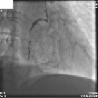
          Unfortunately, right coronary artery (RCA) of a 55-year-old man with unstable angina was spirally dissected from ostium into mid-RCA during manipulation of a guiding catheter and extended to coronary sinus of aortic root (Figure 1). A guidewire could not be introduced into the true lumen. Intravascular ultrasonography-guided wiring could not be attempted due to emergent situation. After initiating medical therapy, chest pain and ST-segment elevation were resolved and hemodynamic status was stabilized. Computerized tomography (CT) performed on the third day showed extended dissection of aortic root was localized at sinus of Valsalva without propagation into ascending aorta (Figure 2). The 7-day follow-up angiography showed collateral flow (Rentrop grade 2) to distal-RCA (Figure 3). Conservative management was maintained for aortic root dissection and delayed percutaneous coronary intervention (PCI) was planned for RCA dissected and totally occluded without surgery. 
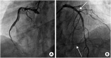
After 3 months, angiography showed no significant change of RCA dissection with slit-like true lumen (Figure 4A). PCI was strugglingly performed from distal-RCA to ostium with 4 stents (Resolute Onyx™) (Figure 4B and C). Then, effort angina was completely improved. Iatrogenic dissection of coronary artery is an uncommon but life-threatening complication.1) When dissection extends to the aortic root and ascending aorta, it should be meticulously evaluated and managed as quickly as possible.2) 3) CT is a helpful and effective tool to evaluate a range of extended dissection flap. If dissection flap is localized and hemodynamic status is stable, conservative management and delayed PCI would be a treatment option for aortocoronary dissection despite acute myocardial infarction.4) 5)

          Related collections

          Most cited references4

          • Record: found
          • Abstract: found
          • Article: not found

          Iatrogenic coronary artery dissections extending into and involving the aortic root.

          We set out to determine the incidence of iatrogenic coronary artery dissection extending into the aorta and to characterize the aortic lesions. We reviewed the data from 43,143 cardiac catheterizations from September 1993 through September 1999 and found 9 coronary artery-aortic dissections for an overall incidence of 0.02%. Four of these patients were undergoing treatment for acute myocardial infarction (AMI) and aortic dissection was more common than for non-AMI patients (0.19% vs. 0.01%, P 40 mm from the coronary os required surgical intervention.
            Bookmark
            • Record: found
            • Abstract: found
            • Article: not found

            Conservative treatment of iatrogenic left main coronary artery dissection: report of two cases.

            Revascularization treatment is mostly recommended for iatrogenic left main coronary artery dissection because of potential for catastrophic sequel, even in case of initial TIMI 3 flow and hemodynamic stability. However, conservative treatment seems to be feasible in asymptomatic and hemodinamically stable patient with low-grade dissection. We report two cases of iatrogenic left main coronary dissection managed conservatively.
              Bookmark
              • Record: found
              • Abstract: found
              • Article: found
              Is Open Access

              Extensive Dissection to the Coronary Sinus of Valsalva During Percutaneous Intervention in Right Coronary Artery—A Case Report and Literature Review

              Severe retrograde dissection extending into the sinus of Valsalva is a rare complication during percutaneous coronary intervention (PCI), but life threatening. There is some literature about this complication, but this particular complication has not been previously reported in China. We present a case of coronary artery dissection during a PCI in which progressively extended retrogradely into the sinus of valsalva, and was successfully treated with stenting without an operation.
                Bookmark

                Author and article information

                Journal
                Korean Circ J
                Korean Circ J
                KCJ
                Korean Circulation Journal
                The Korean Society of Cardiology
                1738-5520
                1738-5555
                April 2019
                07 March 2019
                : 49
                : 4
                : 363-365
                Affiliations
                [1 ]Department of Thoracic and Cardiovascular Surgery, Gil Medical Center, Gachon Cardiovascular Research Institute, Gachon University College of Medicine, Incheon, Korea.
                [2 ]Cardiology Division, Department of Internal Medicine, Gil Medical Center, Gachon Cardiovascular Research Institute, Gachon University College of Medicine, Incheon, Korea.
                Author notes
                Correspondence to Pyung Chun Oh, MD. Cardiology Division, Department of Internal Medicine, Gil Medical Center, Gachon Cardiovascular Research Institute, Gachon University College of Medicine, 21, Namdong-daero 774 beon-gil, Namdong-gu, Incheon 21565, Korea. likemed@ 123456gilhospital.com
                Author information
                https://orcid.org/0000-0002-8538-4511
                https://orcid.org/0000-0003-4590-7178
                https://orcid.org/0000-0002-9955-223X
                Article
                10.4070/kcj.2018.0284
                6428946
                30895759
                342aebe4-24d0-4222-a59b-8dff70a32215
                Copyright © 2019. The Korean Society of Cardiology

                This is an Open Access article distributed under the terms of the Creative Commons Attribution Non-Commercial License ( https://creativecommons.org/licenses/by-nc/4.0) which permits unrestricted noncommercial use, distribution, and reproduction in any medium, provided the original work is properly cited.

                History
                : 16 August 2018
                : 26 November 2018
                : 20 February 2019
                Categories
                Images in Cardiovascular Medicine

                Cardiovascular Medicine
                Cardiovascular Medicine

                Comments

                Comment on this article