22
views
0
recommends
+1 Recommend
0 collections
    0
    shares
      • Record: found
      • Abstract: found
      • Article: found
      Is Open Access

      Endoscopic Management of Peri-Pancreatic Fluid Collections

      review-article

      Read this article at
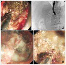
      Bookmark
          There is no author summary for this article yet. Authors can add summaries to their articles on ScienceOpen to make them more accessible to a non-specialist audience.

          Abstract

          In the past decade, there has been a progressive paradigm shift in the management of peri-pancreatic fluid collections after acute pancreatitis. Refinements in the definitions of fluid collections from the updated Atlanta classification have enabled better communication amongst physicians in an effort to formulate optimal treatments. Endoscopic ultrasound (EUS)-guided drainage of pancreatic pseudocysts has emerged as the procedure of choice over surgical cystogastrostomy. The approach provides similar success rates with low complications and better quality of life compared with surgery. However, an endoscopic “step up” approach in the management of pancreatic walled-off necrosis has also been advocated. Both endoscopic and percutaneous drainage routes may be used depending on the anatomical location of the collections. New-generation large diameter EUS-specific stent systems have also recently been described. The device allows precise and effective drainage of the collections and permits endoscopic necrosectomy through the stents.

          Related collections

          Most cited references50

          • Record: found
          • Abstract: found
          • Article: not found

          A clinically based classification system for acute pancreatitis. Summary of the International Symposium on Acute Pancreatitis, Atlanta, Ga, September 11 through 13, 1992.

          E Bradley (1993)
          Acute pancreatitis is a protean disease capable of wide clinical variation, ranging from mild discomfort to apocalyptic prostration. Moreover, the inflammatory process may remain localized in the pancreas, spread to regional tissues, or even involve remote organ systems. This variability in presentation and clinical course has plagued the study and management of acute pancreatitis since its original clinical description. In the absence of accepted definitions for acute pancreatitis and its complications, it has not been possible to devise a clinical classification system useful for case management. Following 3 days of group meetings and open discussions, unanimous consensus on a series of definitions and a clinically based classification system for acute pancreatitis was achieved by a diverse group of 40 international authorities from six medical disciplines and 15 countries. The proposed classification system will be of value to practicing clinicians in the care of individual patients and to academicians seeking to compare interinstitutional data.
            Bookmark
            • Record: found
            • Abstract: found
            • Article: not found

            Clinical evaluation of a novel lumen-apposing metal stent for endosonography-guided pancreatic pseudocyst and gallbladder drainage (with videos).

            Tubular plastic and metal stents have inherent shortcomings when used for transenteric drainage of fluid collections. To evaluate a novel lumen-apposing, self-expandable metal stent for EUS-guided drainage of pancreatic pseudocysts and the gallbladder. Retrospective case series. Tertiary-care academic medical center. This study involved 15 patients (median age 54 years) with symptomatic pancreatic pseudocysts who underwent 12 transgastric and 3 transduodenal pseudocyst drainage procedures. Five patients (median age 69.5 years) with acute cholecystitis underwent 4 cholecystoduodenostomies and 1 cholecystogastostomy. Stent deployment under EUS guidance, passage of an endoscope through the stent lumen for pseudocystoscopy or cholecystoscopy, transenteric endoscopy-guided interventions including biopsy, necrosectomy, and stone removal. Technical and clinical success. All stents were successfully deployed without complication, with a median time to removal of 35 days. All pseudocysts resolved after a single drainage procedure. One stent migrated into the stomach, and the remaining 14 were found to be patent at the time of removal. There was no pseudocyst recurrence during the 11.4-month median follow-up period. One gallbladder stent remains indwelling and fully patent at 12 months. Resolution of acute cholecystitis was observed immediately after stent implantation. No recurrence of symptoms was observed during a median follow-up period of 9 months. Retrospective study, small sample size, lack of control patients. Transenteric drainage of pancreatic pseudocysts and the gallbladder by using a novel, lumen-apposing, metal stent was accomplished with high technical and clinical success in this pilot observational study. Further studies are warranted. Copyright © 2012 American Society for Gastrointestinal Endoscopy. Published by Mosby, Inc. All rights reserved.
              Bookmark
              • Record: found
              • Abstract: found
              • Article: not found

              Acute necrotizing pancreatitis: treatment strategy according to the status of infection.

              To determine benefits of conservative versus surgical treatment in patients with necrotizing pancreatitis. Infection of pancreatic necrosis is the most important risk factor contributing to death in severe acute pancreatitis, and it is generally accepted that infected pancreatic necrosis should be managed surgically. In contrast, the management of sterile pancreatic necrosis accompanied by organ failure is controversial. Recent clinical experience has provided evidence that conservative management of sterile pancreatic necrosis including early antibiotic administration seems promising. A prospective single-center trial evaluated the role of nonsurgical management including early antibiotic treatment in patients with necrotizing pancreatitis. Pancreatic infection, if confirmed by fine-needle aspiration, was considered an indication for surgery, whereas patients without signs of pancreatic infection were treated without surgery. Between January 1994 and June 1999, 204 consecutive patients with acute pancreatitis were recruited. Eighty-six (42%) had necrotizing disease, of whom 57 (66%) had sterile and 29 (34%) infected necrosis. Patients with infected necrosis had more organ failures and a greater extent of necrosis compared with those with sterile necrosis. When early antibiotic treatment was used in all patients with necrotizing pancreatitis (imipenem/cilastatin), the characteristics of pancreatic infection changed to predominantly gram-positive and fungal infections. Fine-needle aspiration showed a sensitivity of 96% for detecting pancreatic infection. The death rate was 1.8% (1/56) in patients with sterile necrosis managed without surgery versus 24% (7/29) in patients with infected necrosis (P <.01). Two patients whose infected necrosis could not be diagnosed in a timely fashion died while receiving nonsurgical treatment. Thus, an intent-to-treat analysis (nonsurgical vs. surgical treatment) revealed a death rate of 5% (3/58) with conservative management versus 21% (6/28) with surgery. These results support nonsurgical management, including early antibiotic treatment, in patients with sterile pancreatic necrosis. Patients with infected necrosis still represent a high-risk group in severe acute pancreatitis, and for them surgical treatment seems preferable.
                Bookmark

                Author and article information

                Journal
                Gut Liver
                Gut Liver
                Gut and Liver
                Editorial Office of Gut and Liver
                1976-2283
                2005-1212
                September 2017
                12 May 2017
                : 11
                : 5
                : 604-611
                Affiliations
                Department of Surgery, Prince of Wales Hospital, The Chinese University of Hong Kong, Hong Kong SAR, China
                Author notes
                Correspondence to: Anthony Yuen Bun Teoh, Department of Surgery, Prince of Wales Hospital, The Chinese University of Hong Kong, 30–32 Ngan Shing Street, Shatin, New Territories, Hong Kong SAR, China, Tel: +852-2632-2627, Fax: +852-2637-7974, E-mail: anthonyteoh@ 123456surgery.cuhk.edu.hk
                Article
                gnl-11-604
                10.5009/gnl16178
                5593321
                28494574
                347f329b-61c5-4254-82ad-09667ea12815
                Copyright © 2017 by The Korean Society of Gastroenterology, the Korean Society of Gastrointestinal Endoscopy, the Korean Society of Neurogastroenterology and Motility, Korean College of Helicobacter and Upper Gastrointestinal Research, Korean Association the Study of Intestinal Diseases, the Korean Association for the Study of the Liver, Korean Pancreatobiliary Association, and Korean Society of Gastrointestinal Cancer.

                This is an Open Access article distributed under the terms of the Creative Commons Attribution Non-Commercial License ( http://creativecommons.org/licenses/by-nc/4.0) which permits unrestricted non-commercial use, distribution, and reproduction in any medium, provided the original work is properly cited.

                History
                : 04 April 2016
                : 10 August 2016
                Categories
                Review

                Gastroenterology & Hepatology
                pancreatic pseudocyst,pancreatic necrosis,endosonography,endoscopic ultrasound-guided pseudocyst drainage,endoscopic necrosectomy

                Comments

                Comment on this article