5
views
0
recommends
+1 Recommend
0 collections
    0
    shares
      • Record: found
      • Abstract: found
      • Article: found
      Is Open Access

      1.5 Tesla Non-ultrashort but Short Echo Time Magnetic Resonance Angiography Describes the Arteries Near a Clipped Cerebral Aneurysm

      case-report

      Read this article at

      Bookmark
          There is no author summary for this article yet. Authors can add summaries to their articles on ScienceOpen to make them more accessible to a non-specialist audience.

          Abstract

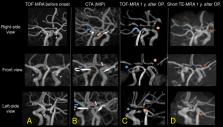
          Cerebral aneurysm and mother artery assessment after clipping is essential to evaluate aneurysm remnant, regrowth, and clip slippage. Usually, cerebral angiography and contrast-enhanced computed tomography angiography (CTA) are used for the evaluation, but they have the side effect of contrast medium and are time-consuming. Time-of-flight magnetic resonance angiography (TOF-MRA) is a non-invasive and fast modality, but clip-induced artifacts limit the signal near the metal clip. Recent ultrashort echo time (UTE)-MRA reduces metal artifacts but its availability is still low worldwide. Therefore, we developed a modified TOF-MRA sequence, named short TE-MRA, using Optima MR 360 1.5T Advance (GE Healthcare Life Sciences, Buckinghamshire, UK). It could describe the artery near the clip using general MRA equipment without recent UTE-MRA technology. We present a subarachnoid hemorrhage patient who underwent short TE-MRA about a year after clipping for the aneurysms at the bilateral internal carotid arteries. Short TE-MRA described the left internal carotid, middle cerebral, and anterior cerebral arteries. The right middle and anterior cerebral arteries were described, but the right internal carotid artery was not. Normal TOF-MRA could not describe them. Without recent UTE-MRA technology, short TE-MRA might be an alternative method for evaluating the artery near the clip. Short TE-MRA can be performed by general MRA equipment with a little time, so it may be helpful until UTE-MRA is widely used. Further research is needed on whether short TE-MRA can describe the aneurysm remnant, regrowth, and clip slippage up to the clinically useful level.

          Related collections

          Most cited references14

          • Record: found
          • Abstract: found
          • Article: not found

          Magnetic resonance imaging near metal implants.

          The desire to apply magnetic resonance imaging (MRI) techniques in the vicinity of embedded metallic hardware is increasing. The soft-tissue contrast available with MR techniques is advantageous in diagnosing complications near an increasing variety of MR-safe metallic hardware. Near such hardware, the spatial encoding mechanisms utilized in conventional MRI methods are often severely compromised. Mitigating these encoding difficulties has been the focus of numerous research investigations over the past two decades. Such approaches include view-angle tilting, short echo-time projection reconstruction acquisitions, single-point imaging, prepolarized MRI, and postprocessing image correction. Various technical advances have also enabled the recent development of two alternative approaches that have shown promising clinical potential. Here, the physical principals and proposed solutions to the problem of MRI near embedded metal are discussed.
            Bookmark
            • Record: found
            • Abstract: found
            • Article: not found

            Volume MR angiography: methods to achieve very short echo times.

            Angiographic displays of cerebral vessels can be generated with single-excitation three-dimensional magnetic resonance imaging. Differentiation of true stenosis from artifactual signal loss, due to dephasing effects from fast or nonconstant blood flow and field inhomogeneities, poses a significant clinical problem that can be largely resolved with the use of very short echo times (TEs). A three-dimensional imaging technique was developed that allows TEs of 3.1 msec without and 4.5 msec with first-order flow compensation gradients. The short TEs were achieved with short asymmetric radio-frequency pulses, gradients of minimal duration, and fractional echoes. Significantly improved images of normal tortuous vessels with fast flow were obtained. With this method, accuracy in depicting the vessel lumen and confidence in the findings were markedly increased.
              Bookmark
              • Record: found
              • Abstract: found
              • Article: not found

              Magnetic resonance: an introduction to ultrashort TE (UTE) imaging.

              The background underpinning the clinical use of ultrashort echo-time (UTE) pulse sequences for imaging tissues or tissue components with short T2s is reviewed. Tissues properties are discussed, and tissues are divided into those with a majority of short T2 relaxation components and those with a minority. Features of the basic physics relevant to UTE imaging are described including the fact that when the radiofrequency pulse duration is of the order T2, rotation of tissue magnetization into the transverse plane is incomplete. Consequences of the broad line-width of short T2 components are also discussed including their partial saturation by off-resonance fat suppression pulses as well as multislice and multiecho imaging. The need for rapid data acquisition of the order T2 is explained. The basic UTE pulse sequence with its half excitation pulse and radial imaging from the center of k-space is described together with options that suppress fat and/or long T2 components. Image interpretation is discussed. Clinical features of the imaging of cortical bone, tendons, ligaments, menisci, and periosteum as well as brain, liver, and spine are illustrated. Short T2 components in all of these tissues may show high signals. Possible future developments are outlined as are technical limitations.
                Bookmark

                Author and article information

                Journal
                Cureus
                Cureus
                2168-8184
                Cureus
                Cureus (Palo Alto (CA) )
                2168-8184
                25 July 2021
                July 2021
                : 13
                : 7
                : e16611
                Affiliations
                [1 ] Department of Radiological Technology, Itoigawa General Hospital, Itoigawa, JPN
                [2 ] Department of Neurosurgery, Itoigawa General Hospital, Itoigawa, JPN
                Author notes
                Article
                10.7759/cureus.16611
                8383129
                3510524a-c4dd-4156-a0bf-84ad30131445
                Copyright © 2021, Higo et al.

                This is an open access article distributed under the terms of the Creative Commons Attribution License, which permits unrestricted use, distribution, and reproduction in any medium, provided the original author and source are credited.

                History
                : 25 July 2021
                Categories
                Neurology
                Radiology
                Neurosurgery

                cerebral aneurysm,clipping,time-of-flight magnetic resonance angiography (tof-mra),ultrashort echo time magnetic resonance angiography (ute-mra),less invasive,magnetic resonance imaging,clip artifact

                Comments

                Comment on this article