32
views
0
recommends
+1 Recommend
0 collections
    0
    shares
      • Record: found
      • Abstract: found
      • Article: found
      Is Open Access

      Ganoderma lucidum polysaccharide peptide prevents renal ischemia reperfusion injury via counteracting oxidative stress

      research-article

      Read this article at

      Bookmark
          There is no author summary for this article yet. Authors can add summaries to their articles on ScienceOpen to make them more accessible to a non-specialist audience.

          Abstract

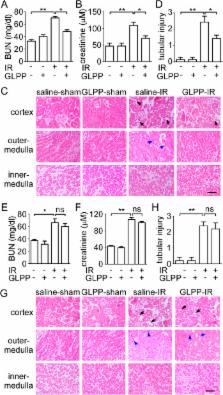
          Ganoderma lucidum polysaccharide peptide (GLPP) scavenges oxygen free radicals that are a key factor in the pathogenesis of renal ischemia reperfusion injury (RIRI). The aim of this study was to determine whether GLPP could attenuate RIRI by counteracting the oxidative stress. The mechanism involved was assessed by an in vivo mouse RIRI model and an in vitro hypoxia/reoxygenation model, and tunicamycin-stimulated NRK-52E cells were used to explore the GLPP-mediated alleviation of ER stress. Experimental results showed that renal dysfunction and morphological damage were reduced in GLPP-treated group. The imbalance of redox status was reversed and production of ROS was reduced by GLPP. RIRI-induced mitochondrial- and ER stress-dependent apoptosis were dramatically inhibited in GLPP-treated group. Intriguingly, JNK activation in the kidney with RIRI or hypoxia/reoxygenation was inhibited by GLPP. These results suggest that the protective effect of GLPP against RIRI may be due to reducing oxidative stress, alleviating the mitochondrial and ER stress-dependent apoptosis caused by excessive ROS.

          Related collections

          Most cited references43

          • Record: found
          • Abstract: found
          • Article: not found

          Induction of apoptosis by ASK1, a mammalian MAPKKK that activates SAPK/JNK and p38 signaling pathways.

          Mitogen-activated protein (MAP) kinase cascades are activated in response to various extracellular stimuli, including growth factors and environmental stresses. A MAP kinase kinase kinase (MAPKKK), termed ASK1, was identified that activated two different subgroups of MAP kinase kinases (MAPKK), SEK1 (or MKK4) and MKK3/MAPKK6 (or MKK6), which in turn activated stress-activated protein kinase (SAPK, also known as JNK; c-Jun amino-terminal kinase) and p38 subgroups of MAP kinases, respectively. Overexpression of ASK1 induced apoptotic cell death, and ASK1 was activated in cells treated with tumor necrosis factor-alpha (TNF-alpha). Moreover, TNF-alpha-induced apoptosis was inhibited by a catalytically inactive form of ASK1. ASK1 may be a key element in the mechanism of stress- and cytokine-induced apoptosis.
            Bookmark
            • Record: found
            • Abstract: found
            • Article: not found

            Pathophysiology of ischemic acute kidney injury.

            Acute kidney injury (AKI) as a consequence of ischemia is a common clinical event leading to unacceptably high morbidity and mortality, development of chronic kidney disease (CKD), and transition from pre-existing CKD to end-stage renal disease. Data indicate a close interaction between the many cell types involved in the pathophysiology of ischemic AKI, which has critical implications for the treatment of this condition. Inflammation seems to be the common factor that links the various cell types involved in this process. In this Review, we describe the interactions between these cells and their response to injury following ischemia. We relate these events to patients who are at high risk of AKI, and highlight the characteristics that might predispose these patients to injury. We also discuss how therapy targeting specific cell types can minimize the initial and subsequent injury following ischemia, thereby limiting the extent of acute changes and, hopefully, long-term structural and functional alterations to the kidney.
              Bookmark
              • Record: found
              • Abstract: found
              • Article: not found

              Regulation of mitochondrial dynamics in acute kidney injury in cell culture and rodent models.

              The mechanism of mitochondrial damage, a key contributor to renal tubular cell death during acute kidney injury, remains largely unknown. Here, we have demonstrated a striking morphological change of mitochondria in experimental models of renal ischemia/reperfusion and cisplatin-induced nephrotoxicity. This change contributed to mitochondrial outer membrane permeabilization, release of apoptogenic factors, and consequent apoptosis. Following either ATP depletion or cisplatin treatment of rat renal tubular cells, mitochondrial fragmentation was observed prior to cytochrome c release and apoptosis. This mitochondrial fragmentation was inhibited by Bcl2 but not by caspase inhibitors. Dynamin-related protein 1 (Drp1), a critical mitochondrial fission protein, translocated to mitochondria early during tubular cell injury, and both siRNA knockdown of Drp1 and expression of a dominant-negative Drp1 attenuated mitochondrial fragmentation, cytochrome c release, caspase activation, and apoptosis. Further in vivo analysis revealed that mitochondrial fragmentation also occurred in proximal tubular cells in mice during renal ischemia/reperfusion and cisplatin-induced nephrotoxicity. Notably, both tubular cell apoptosis and acute kidney injury were attenuated by mdivi-1, a newly identified pharmacological inhibitor of Drp1. This study demonstrates a rapid regulation of mitochondrial dynamics during acute kidney injury and identifies mitochondrial fragmentation as what we believe to be a novel mechanism contributing to mitochondrial damage and apoptosis in vivo in mouse models of disease.
                Bookmark

                Author and article information

                Journal
                Sci Rep
                Sci Rep
                Scientific Reports
                Nature Publishing Group
                2045-2322
                25 November 2015
                2015
                : 5
                : 16910
                Affiliations
                [1 ]State Key Laboratory of Natural and Biomimetic Drugs, Department of Pharmacology, School of Basic Medical Sciences, Peking University , Beijing, China
                [2 ]Fuzhou Institute of Green Valley Bio-Pharm Technology , Fuzhou, China
                [3 ]JUNCAO Technology Research Institute, Fujian Agriculture and Forestry University , Fuzhou, China
                Author notes
                Article
                srep16910
                10.1038/srep16910
                4658483
                26603550
                351bd04a-fc43-4704-99a1-3842cad747f9
                Copyright © 2015, Macmillan Publishers Limited

                This work is licensed under a Creative Commons Attribution 4.0 International License. The images or other third party material in this article are included in the article’s Creative Commons license, unless indicated otherwise in the credit line; if the material is not included under the Creative Commons license, users will need to obtain permission from the license holder to reproduce the material. To view a copy of this license, visit http://creativecommons.org/licenses/by/4.0/

                History
                : 03 July 2015
                : 21 October 2015
                Categories
                Article

                Uncategorized
                Uncategorized

                Comments

                Comment on this article