Inviting an author to review:
Find an author and click ‘Invite to review selected article’ near their name.
Search for authorsSearch for similar articles
4
views
0
recommends
+1 Recommend
0 collections
    0
    shares
      • Record: found
      • Abstract: found
      • Article: found
      Is Open Access

      Nano-seq analysis reveals different functional tendency between exosomes and microvesicles derived from hUMSC

      research-article

      Read this article at

      Bookmark
          There is no author summary for this article yet. Authors can add summaries to their articles on ScienceOpen to make them more accessible to a non-specialist audience.

          Abstract

          Background

          Extracellular vesicles (EVs) from human umbilical cord mesenchymal stem cells (hUMSCs) are widely considered to be the best mediators for cell-free therapy. An understanding of their composition, especially RNA, is particularly important for the safe and precise application of EVs. Up to date, the knowledge of their RNA components is limited to NGS sequencing and cannot provide a comprehensive transcriptomic landscape, especially the long and full-length transcripts. Our study first focused on the transcriptomic profile of hUMSC-EVs based on nanopore sequencing.

          Methods

          In this study, different EV subtypes (exosomes and microvesicles) derived from hUMSCs were isolated and identified by density gradient centrifugation. Subsequently, the realistic long transcriptomic profile in different subtypes of hUMSC-EVs was systematically compared by nanopore sequencing and bioinformatic analysis.

          Results

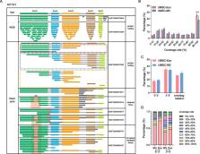
          Abundant transcript variants were identified in EVs by nanopore sequencing, 69.34% of which transcripts were fragmented. A series of full-length and long transcripts was also observed and showed a significantly higher proportion of intact or near-complete transcripts in exosomes than that in microvesicles derived from hUMSCs. Although the composition of RNA biotypes transported by different EV subtypes was similar, the distribution of transcripts and genes revealed the inter-heterogeneity and intra-stability between exosomes and microvesicles. Further, 85 different expressed transcripts (56 genes) and 7 fusion genes were identified. Pathway enrichment analysis showed that upregulated-expressed genes in microvesicles were mainly enriched in multiple neurodegenerative diseases, while upregulated-expressed genes in exosomes were mainly enriched in neutrophil extracellular trap formation, suggesting different functional tendencies of EV subtypes.

          Conclusions

          This study provides a novel understanding of different types of hUMSC-EVs, which not only suggests different transcriptome sorting mechanisms between exosomes and microvesicles, but also shows that different EV subtypes from the same source have different physiological functions, suggesting distinct clinical application prospects.

          Supplementary Information

          The online version contains supplementary material available at 10.1186/s13287-023-03491-5.

          Related collections

          Most cited references38

          • Record: found
          • Abstract: found
          • Article: not found

          Exosome-mediated transfer of mRNAs and microRNAs is a novel mechanism of genetic exchange between cells.

          Exosomes are vesicles of endocytic origin released by many cells. These vesicles can mediate communication between cells, facilitating processes such as antigen presentation. Here, we show that exosomes from a mouse and a human mast cell line (MC/9 and HMC-1, respectively), as well as primary bone marrow-derived mouse mast cells, contain RNA. Microarray assessments revealed the presence of mRNA from approximately 1300 genes, many of which are not present in the cytoplasm of the donor cell. In vitro translation proved that the exosome mRNAs were functional. Quality control RNA analysis of total RNA derived from exosomes also revealed presence of small RNAs, including microRNAs. The RNA from mast cell exosomes is transferable to other mouse and human mast cells. After transfer of mouse exosomal RNA to human mast cells, new mouse proteins were found in the recipient cells, indicating that transferred exosomal mRNA can be translated after entering another cell. In summary, we show that exosomes contain both mRNA and microRNA, which can be delivered to another cell, and can be functional in this new location. We propose that this RNA is called "exosomal shuttle RNA" (esRNA).
            Bookmark
            • Record: found
            • Abstract: found
            • Article: found
            Is Open Access

            Exosome and Exosomal MicroRNA: Trafficking, Sorting, and Function

            Exosomes are 40–100 nm nano-sized vesicles that are released from many cell types into the extracellular space. Such vesicles are widely distributed in various body fluids. Recently, mRNAs and microRNAs (miRNAs) have been identified in exosomes, which can be taken up by neighboring or distant cells and subsequently modulate recipient cells. This suggests an active sorting mechanism of exosomal miRNAs, since the miRNA profiles of exosomes may differ from those of the parent cells. Exosomal miRNAs play an important role in disease progression, and can stimulate angiogenesis and facilitate metastasis in cancers. In this review, we will introduce the origin and the trafficking of exosomes between cells, display current research on the sorting mechanism of exosomal miRNAs, and briefly describe how exosomes and their miRNAs function in recipient cells. Finally, we will discuss the potential applications of these miRNA-containing vesicles in clinical settings.
              Bookmark
              • Record: found
              • Abstract: found
              • Article: not found
              Is Open Access

              Mesenchymal Stem Cell-derived Extracellular Vesicles: Toward Cell-free Therapeutic Applications.

              Mesenchymal stem (stromal) cells (MSCs) are multipotent cells with the ability to differentiate into several cell types, thus serving as a cell reservoir for regenerative medicine. Much of the current interest in therapeutic application of MSCs to various disease settings can be linked to their immunosuppressive and anti-inflammatory properties. One of the key mechanisms of MSC anti-inflammatory effects is the secretion of soluble factors with paracrine actions. Recently it has emerged that the paracrine functions of MSCs could, at least in part, be mediated by extracellular vesicles (EVs). EVs are predominantly released from the endosomal compartment and contain a cargo that includes miRNA, mRNA, and proteins from their cells of origin. Recent animal model-based studies suggest that EVs have significant potential as a novel alternative to whole cell therapies. Compared to their parent cells, EVs may have a superior safety profile and can be safely stored without losing function. In this article, we review current knowledge related to the potential use of MSC-derived EVs in various diseases and discuss the promising future for EVs as an alternative, cell-free therapy.
                Bookmark

                Author and article information

                Contributors
                wangyuesmmu@126.com
                Journal
                Stem Cell Res Ther
                Stem Cell Res Ther
                Stem Cell Research & Therapy
                BioMed Central (London )
                1757-6512
                25 September 2023
                25 September 2023
                2023
                : 14
                : 272
                Affiliations
                [1 ]GRID grid.73113.37, ISNI 0000 0004 0369 1660, Department of Precision Medicine, Translational Medicine Research Center, , Naval Medical University, ; Shanghai, People’s Republic of China
                [2 ]GRID grid.73113.37, ISNI 0000 0004 0369 1660, Department of Stem Cell and Regeneration Medicine, Translational Medicine Research Center, , Naval Medical University, ; Xiangyin Road 800, Shanghai, People’s Republic of China
                [3 ]GRID grid.73113.37, ISNI 0000 0004 0369 1660, Department of Histology and Embryology, Basic Medicine Collage, , Naval Medical University, ; Shanghai, People’s Republic of China
                [4 ]Department of Plastic and Reconstructive Surgery, Secondary Affiliated Hospital of Naval Medical University, ( https://ror.org/01wn7w598) Shanghai, People’s Republic of China
                [5 ]Medical College of Guangxi University, ( https://ror.org/02c9qn167) Nanning, People’s Republic of China
                [6 ]Shanghai Key Laboratory of Cell Engineering, Shanghai, People’s Republic of China
                [7 ]Shanghai Institute of Stem Cell Research and Clinical Translation, Shanghai, People’s Republic of China
                Article
                3491
                10.1186/s13287-023-03491-5
                10521478
                37749641
                37bf31e1-8fef-428f-b89a-fd98b37b54bd
                © BioMed Central Ltd., part of Springer Nature 2023

                Open Access This article is licensed under a Creative Commons Attribution 4.0 International License, which permits use, sharing, adaptation, distribution and reproduction in any medium or format, as long as you give appropriate credit to the original author(s) and the source, provide a link to the Creative Commons licence, and indicate if changes were made. The images or other third party material in this article are included in the article's Creative Commons licence, unless indicated otherwise in a credit line to the material. If material is not included in the article's Creative Commons licence and your intended use is not permitted by statutory regulation or exceeds the permitted use, you will need to obtain permission directly from the copyright holder. To view a copy of this licence, visit http://creativecommons.org/licenses/by/4.0/. The Creative Commons Public Domain Dedication waiver ( http://creativecommons.org/publicdomain/zero/1.0/) applies to the data made available in this article, unless otherwise stated in a credit line to the data.

                History
                : 6 September 2022
                : 7 September 2023
                Funding
                Funded by: National key research and development program
                Award ID: Grant No. 2018YFA0108301
                Award Recipient :
                Funded by: FundRef http://dx.doi.org/10.13039/501100001809, National Natural Science Foundation of China;
                Award ID: Grant No. 31971109
                Award Recipient :
                Funded by: Shanghai Key Laboratory of Cell Engineering
                Award ID: NO. :14DZ2272300
                Award Recipient :
                Funded by: Shanghai Sailing Program
                Award ID: NO.:20YF1458100
                Award Recipient :
                Funded by: Shanghai Biomedical Science and Technology Project
                Award ID: Grant No. 22S11901600
                Award Recipient :
                Funded by: Technical product research project
                Award ID: Grant No. AWS18C001
                Award Recipient :
                Funded by: Naval Medical University and University of Shanghai for Science and Technology Joint Projects
                Award ID: 2020-RZ04
                Award Recipient :
                Funded by: Peak Disciplines (Type IV) of Institutions of Higher Learning in Shanghai
                Categories
                Research
                Custom metadata
                © BioMed Central Ltd., part of Springer Nature 2023

                Molecular medicine
                extracellular vesicles subtypes,nanopore sequencing,transcriptomic landscape

                Comments

                Comment on this article