38
views
0
recommends
+1 Recommend
0 collections
    0
    shares
      • Record: found
      • Abstract: found
      • Article: found
      Is Open Access

      Impaired Cerebral Autoregulation Is Associated with Brain Atrophy and Worse Functional Status in Chronic Ischemic Stroke

      research-article

      Read this article at

      Bookmark
          There is no author summary for this article yet. Authors can add summaries to their articles on ScienceOpen to make them more accessible to a non-specialist audience.

          Abstract

          Dynamic cerebral autoregulation (dCA) is impaired following stroke. However, the relationship between dCA, brain atrophy, and functional outcomes following stroke remains unclear. In this study, we aimed to determine whether impairment of dCA is associated with atrophy in specific regions or globally, thereby affecting daily functions in stroke patients.

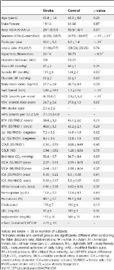
          We performed a retrospective analysis of 33 subjects with chronic infarctions in the middle cerebral artery territory, and 109 age-matched non-stroke subjects. dCA was assessed via the phase relationship between arterial blood pressure and cerebral blood flow velocity. Brain tissue volumes were quantified from MRI. Functional status was assessed by gait speed, instrumental activities of daily living (IADL), modified Rankin Scale, and NIH Stroke Score.

          Compared to the non-stroke group, stroke subjects showed degraded dCA bilaterally, and showed gray matter atrophy in the frontal, parietal and temporal lobes ipsilateral to infarct. In stroke subjects, better dCA was associated with less temporal lobe gray matter atrophy on the infracted side (  = 0.029), faster gait speed (  = 0.018) and lower IADL score ( 0.002). Our results indicate that better dynamic cerebral perfusion regulation is associated with less atrophy and better long-term functional status in older adults with chronic ischemic infarctions.

          Related collections

          Most cited references34

          • Record: found
          • Abstract: found
          • Article: not found

          Cerebral autoregulation.

          Autoregulation of blood flow denotes the intrinsic ability of an organ or a vascular bed to maintain a constant perfusion in the face of blood pressure changes. Alternatively, autoregulation can be defined in terms of vascular resistance changes or simply arteriolar caliber changes as blood pressure or perfusion pressure varies. While known in almost any vascular bed, autoregulation and its disturbance by disease has attracted particular attention in the cerebrovascular field. The basic mechanism of autoregulation of cerebral blood flow (CBF) is controversial. Most likely, the autoregulatory vessel caliber changes are mediated by an interplay between myogenic and metabolic mechanisms. Influence of perivascular nerves and most recently the vascular endothelium has also been the subject of intense investigation. CBF autoregulation typically operates between mean blood pressures of the order of 60 and 150 mm Hg. These limits are not entirely fixed but can be modulated by sympathetic nervous activity, the vascular renin-angiotensin system, and any factor (notably changes in arterial carbon dioxide tension) that decreases or increases CBF. Disease states of the brain may impair or abolish CBF autoregulation. Thus, autoregulation is lost in severe head injury or acute ischemic stroke, leaving surviving brain tissue unprotected against the potentially harmful effect of blood pressure changes. Likewise, autoregulation may be lost in the surroundings of a space-occupying brain lesion, be it a tumor or a hematoma. In many such disease states, autoregulation may be regained by hyperventilatory hypocapnia. Autoregulation may also be impaired in neonatal brain asphyxia and infections of the central nervous system, but appears to be intact in spreading depression and migraine, despite impairment of chemical and metabolic control of CBF. In chronic hypertension, the limits of autoregulation are shifted toward high blood pressure. Acute hypertensive encephalopathy, on the other hand, is thought to be due to autoregulatory failure at very high pressure. In long-term diabetes mellitus there may be chronic impairment of CBF autoregulation, probably due to diabetic microangiopathy.
            Bookmark
            • Record: found
            • Abstract: found
            • Article: not found

            Telmisartan to prevent recurrent stroke and cardiovascular events.

            Prolonged lowering of blood pressure after a stroke reduces the risk of recurrent stroke. In addition, inhibition of the renin-angiotensin system in high-risk patients reduces the rate of subsequent cardiovascular events, including stroke. However, the effect of lowering of blood pressure with a renin-angiotensin system inhibitor soon after a stroke has not been clearly established. We evaluated the effects of therapy with an angiotensin-receptor blocker, telmisartan, initiated early after a stroke. In a multicenter trial involving 20,332 patients who recently had an ischemic stroke, we randomly assigned 10,146 to receive telmisartan (80 mg daily) and 10,186 to receive placebo. The primary outcome was recurrent stroke. Secondary outcomes were major cardiovascular events (death from cardiovascular causes, recurrent stroke, myocardial infarction, or new or worsening heart failure) and new-onset diabetes. The median interval from stroke to randomization was 15 days. During a mean follow-up of 2.5 years, the mean blood pressure was 3.8/2.0 mm Hg lower in the telmisartan group than in the placebo group. A total of 880 patients (8.7%) in the telmisartan group and 934 patients (9.2%) in the placebo group had a subsequent stroke (hazard ratio in the telmisartan group, 0.95; 95% confidence interval [CI], 0.86 to 1.04; P=0.23). Major cardiovascular events occurred in 1367 patients (13.5%) in the telmisartan group and 1463 patients (14.4%) in the placebo group (hazard ratio, 0.94; 95% CI, 0.87 to 1.01; P=0.11). New-onset diabetes occurred in 1.7% of the telmisartan group and 2.1% of the placebo group (hazard ratio, 0.82; 95% CI, 0.65 to 1.04; P=0.10). Therapy with telmisartan initiated soon after an ischemic stroke and continued for 2.5 years did not significantly lower the rate of recurrent stroke, major cardiovascular events, or diabetes. (ClinicalTrials.gov number, NCT00153062.) 2008 Massachusetts Medical Society
              Bookmark
              • Record: found
              • Abstract: found
              • Article: not found

              Autonomic neural control of dynamic cerebral autoregulation in humans.

              The purpose of the present study was to determine the role of autonomic neural control of dynamic cerebral autoregulation in humans. We measured arterial pressure and cerebral blood flow (CBF) velocity in 12 healthy subjects (aged 29+/-6 years) before and after ganglion blockade with trimethaphan. CBF velocity was measured in the middle cerebral artery using transcranial Doppler. The magnitude of spontaneous changes in mean blood pressure and CBF velocity were quantified by spectral analysis. The transfer function gain, phase, and coherence between these variables were estimated to quantify dynamic cerebral autoregulation. After ganglion blockade, systolic and pulse pressure decreased significantly by 13% and 26%, respectively. CBF velocity decreased by 6% (P<0.05). In the very low frequency range (0.02 to 0.07 Hz), mean blood pressure variability decreased significantly (by 82%), while CBF velocity variability persisted. Thus, transfer function gain increased by 81%. In addition, the phase lead of CBF velocity to arterial pressure diminished. These changes in transfer function gain and phase persisted despite restoration of arterial pressure by infusion of phenylephrine and normalization of mean blood pressure variability by oscillatory lower body negative pressure. These data suggest that dynamic cerebral autoregulation is altered by ganglion blockade. We speculate that autonomic neural control of the cerebral circulation is tonically active and likely plays a significant role in the regulation of beat-to-beat CBF in humans.
                Bookmark

                Author and article information

                Contributors
                Role: Editor
                Journal
                PLoS One
                PLoS ONE
                plos
                plosone
                PLoS ONE
                Public Library of Science (San Francisco, USA )
                1932-6203
                2012
                11 October 2012
                : 7
                : 10
                : e46794
                Affiliations
                [1 ]Biomathematics Program, Department of Mathematics, North Carolina State University, Raleigh, North Carolina, United States of America
                [2 ]Division of Sleep Medicine, Brigham and Women's Hospital, Harvard Medical School, Boston, Massachusetts, United States of America
                [3 ]The Research Center for Adaptive Data Analysis/Center for Dynamical Biomarker and Translational Medicine, National Central University, Chungli, Taiwan, Republic of China
                [4 ]Department of Neurology, Beth Israel Deaconess Medical Center, Harvard Medical School, Boston, Massachusetts, United States of America
                [5 ]Division of Gerontology, Beth Israel Deaconess Medical Center, Harvard Medical School, Boston, Massachusetts, United States of America
                University of Queensland, Australia
                Author notes

                Competing Interests: The authors have declared that no competing interests exist.

                Conceived and designed the experiments: VN KH. Performed the experiments: VN. Analyzed the data: MCA KH VN. Contributed reagents/materials/analysis tools: KH MTL. Wrote the paper: MCA KH VN. Provided expert interpretation of data analysis and edited the manuscript: MS MSO.

                Article
                PONE-D-12-08015
                10.1371/journal.pone.0046794
                3469603
                23071639
                38e5656c-6fd4-4ad2-8b6e-e68e80b1f482
                Copyright @ 2012

                This is an open-access article distributed under the terms of the Creative Commons Attribution License, which permits unrestricted use, distribution, and reproduction in any medium, provided the original author and source are credited.

                History
                : 19 March 2012
                : 7 September 2012
                Page count
                Pages: 9
                Funding
                This study was supported by The National Institutes of Health - National Institute of Neurological Disorders and Stroke (NIH - NINDS) ( www.ninds.nih.gov/) (R01-NS045745), The National Institutes of Health - National Institute of Neurological Disorders and Stroke (NIH - NINDS), The National Institutes of Health - National Institute of Neurological Disorders and Stroke - The Small Business Technology Transfer (NIH - NINDS - STTR) (1R41NS053128-01A2), The National Institutes of Health - National Institute of Aging (NIH-NIA) (1R01-AG0287601A2), (1P30AG028717-01A2), American Diabetes Association (1-06-CR-25), and The National Institutes of Health - National Institute of Aging (NIH-NIA) (OAIC1P30AG028717-01A2). MCA and MSO were supported in part by the National Science Foundation/Division of Mathematical Sciences-0616597 and Kirschstein-National Research Service Award 5 T32 AG023480 - 05 grants. MCA also received support from the Center for Quantitative Sciences in Biology at North Carolina State University. KH was supported by The National Institutes of Health - The National Heart, Lung, and Blood Institute (NIH - NHLBI) K99HL102241, NIH-R00HL102241, P30AG028717, and the KL2 Medical Research Investigator Training grant (5 KL2 RR025757-02) of Harvard Catalyst, The Harvard Clinical and Translational Science Center (Award #UL1 RR 025758 and financial contributions from Harvard University and its affiliated academic health care centers). MS receives grant support from the The National Institutes of Health - National Institute of Neurological Disorders and Stroke (NIH/NINDS) (RO1 NS057127). VN and KH were also supported by Grant Number UL1 RR025758-Harvard Clinical and Translational Science Center, from the National Center for Research Resources and a Beth Israel Deaconess Medical Center, Clinical Research Center Grant (MO1-RR01302). MTL was supported by National Science Council (NSC) (Taiwan, ROC) grants 100-2221-E-008-008-MY2 and 100-2911-I-008-001. The funders had no role in study design, data collection and analysis, decision to publish, or preparation of the manuscript.
                Categories
                Research Article
                Engineering
                Signal Processing
                Signal Filtering
                Medicine
                Cardiovascular
                Stroke
                Neurology
                Cerebrovascular Diseases
                Ischemic Stroke
                Autonomic Nervous System
                Neurorehabilitation and Trauma
                Radiology
                Diagnostic Radiology
                Magnetic Resonance Imaging

                Uncategorized
                Uncategorized

                Comments

                Comment on this article