6
views
0
recommends
+1 Recommend
0 collections
    0
    shares
      • Record: found
      • Abstract: found
      • Article: found
      Is Open Access

      Artifact Reduction in Simultaneous EEG-fMRI: A Systematic Review of Methods and Contemporary Usage

      systematic-review

      Read this article at

      Bookmark
          There is no author summary for this article yet. Authors can add summaries to their articles on ScienceOpen to make them more accessible to a non-specialist audience.

          Abstract

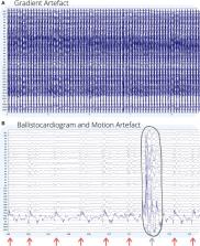
          Simultaneous electroencephalography-functional MRI (EEG-fMRI) is a technique that combines temporal (largely from EEG) and spatial (largely from fMRI) indicators of brain dynamics. It is useful for understanding neuronal activity during many different event types, including spontaneous epileptic discharges, the activity of sleep stages, and activity evoked by external stimuli and decision-making tasks. However, EEG recorded during fMRI is subject to imaging, pulse, environment and motion artifact, causing noise many times greater than the neuronal signals of interest. Therefore, artifact removal methods are essential to ensure that artifacts are accurately removed, and EEG of interest is retained. This paper presents a systematic review of methods for artifact reduction in simultaneous EEG-fMRI from literature published since 1998, and an additional systematic review of EEG-fMRI studies published since 2016. The aim of the first review is to distill the literature into clear guidelines for use of simultaneous EEG-fMRI artifact reduction methods, and the aim of the second review is to determine the prevalence of artifact reduction method use in contemporary studies. We find that there are many published artifact reduction techniques available, including hardware, model based, and data-driven methods, but there are few studies published that adequately compare these methods. In contrast, recent EEG-fMRI studies show overwhelming use of just one or two artifact reduction methods based on literature published 15–20 years ago, with newer methods rarely gaining use outside the group that developed them. Surprisingly, almost 15% of EEG-fMRI studies published since 2016 fail to adequately describe the methods of artifact reduction utilized. We recommend minimum standards for reporting artifact reduction techniques in simultaneous EEG-fMRI studies and suggest that more needs to be done to make new artifact reduction techniques more accessible for the researchers and clinicians using simultaneous EEG-fMRI.

          Related collections

          Most cited references145

          • Record: found
          • Abstract: found
          • Article: not found

          Preferred reporting items for systematic reviews and meta-analyses: the PRISMA statement

          David Moher and colleagues introduce PRISMA, an update of the QUOROM guidelines for reporting systematic reviews and meta-analyses
            Bookmark
            • Record: found
            • Abstract: not found
            • Article: not found

            A real-time QRS detection algorithm.

              Bookmark
              • Record: found
              • Abstract: found
              • Article: not found

              Identification of EEG events in the MR scanner: the problem of pulse artifact and a method for its subtraction.

              Triggering functional MRI (fMRI) image acquisition immediately after an EEG event can provide information on the location of the event generator. However, EEG artifact associated with pulsatile blood flow in a subject inside the scanner may obscure EEG events. This pulse artifact (PA) has been widely recognized as a significant problem, although its characteristics are unpredictable. We have investigated the amplitude, distribution on the scalp, and frequency of occurrence of this artifact. This showed large interindividual variations in amplitude, although PA is normally largest in the frontal region. In five of six subjects, PA was greater than 50 microV in at least one of the temporal, parasagittal, and central channels analyzed. Therefore, we developed and validated a method for removing PA. This subtracts an averaged PA waveform calculated for each electrode during the previous 10 s. Particular attention has been given to reliable ECG peak detection and ensuring that the average PA waveform is free of other EEG artifacts. Comparison of frequency spectra for EEG recorded outside and inside the scanner, with and without PA subtraction, showed a clear reduction in artifact after PA subtraction for all four frequency ranges analyzed. As further validation, lateralized epileptiform spikes were added to recordings from inside and outside the scanner: PA subtraction significantly increased the proportion of these spikes that were correctly identified and decreased the number of false spike detections. We conclude that in some subjects, EEG/fMRI studies will be feasible only using PA subtraction. Copyright 1998 Academic Press.
                Bookmark

                Author and article information

                Contributors
                Journal
                Front Neurol
                Front Neurol
                Front. Neurol.
                Frontiers in Neurology
                Frontiers Media S.A.
                1664-2295
                11 March 2021
                2021
                : 12
                : 622719
                Affiliations
                [1] 1Florey Department of Neuroscience and Mental Health, The University of Melbourne , Melbourne, VIC, Australia
                [2] 2Florey Institute of Neuroscience and Mental Health , Melbourne, VIC, Australia
                [3] 3Department of Medicine (Austin Health), The University of Melbourne , Melbourne, VIC, Australia
                Author notes

                Edited by: Fernando Cendes, State University of Campinas, Brazil

                Reviewed by: Li Dong, University of Electronic Science and Technology of China, China; Jean Gotman, McGill University, Canada

                *Correspondence: David F. Abbott david.abbott@ 123456florey.edu.au

                This article was submitted to Applied Neuroimaging, a section of the journal Frontiers in Neurology

                Article
                10.3389/fneur.2021.622719
                7991907
                33776886
                38f18b0f-554e-4882-b1d3-27628fa99ca0
                Copyright © 2021 Bullock, Jackson and Abbott.

                This is an open-access article distributed under the terms of the Creative Commons Attribution License (CC BY). The use, distribution or reproduction in other forums is permitted, provided the original author(s) and the copyright owner(s) are credited and that the original publication in this journal is cited, in accordance with accepted academic practice. No use, distribution or reproduction is permitted which does not comply with these terms.

                History
                : 29 October 2020
                : 29 January 2021
                Page count
                Figures: 7, Tables: 1, Equations: 0, References: 146, Pages: 21, Words: 16881
                Categories
                Neurology
                Systematic Review

                Neurology
                simultaneous eeg-fmri,artifact,motion,ballistocardiogram,electroencephalography,bold
                Neurology
                simultaneous eeg-fmri, artifact, motion, ballistocardiogram, electroencephalography, bold

                Comments

                Comment on this article