10
views
0
recommends
+1 Recommend
0 collections
    0
    shares
      • Record: found
      • Abstract: found
      • Article: found
      Is Open Access

      Fibroblast-induced switching to the mesenchymal-like phenotype and PI3K/mTOR signaling protects melanoma cells from BRAF inhibitors

      research-article

      Read this article at

      Bookmark
          There is no author summary for this article yet. Authors can add summaries to their articles on ScienceOpen to make them more accessible to a non-specialist audience.

          Abstract

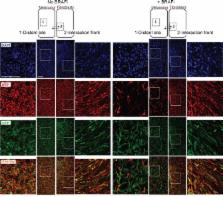
          The knowledge on how tumor-associated stroma influences efficacy of anti-cancer therapy just started to emerge. Here we show that lung fibroblasts reduce melanoma sensitivity to the BRAF inhibitor (BRAFi) vemurafenib only if the two cell types are in close proximity. In the presence of fibroblasts, the adjacent melanoma cells acquire de-differentiated mesenchymal-like phenotype. Upon treatment with BRAFi, such melanoma cells maintain high levels of phospho ribosomal protein S6 (pS6), i.e. active mTOR signaling, which is suppressed in the BRAFi sensitive cells without stromal contacts. Inhibitors of PI3K/mTOR in combination with BRAFi eradicate pS6 high cell subpopulations and potentiate anti-cancer effects in melanoma protected by the fibroblasts. mTOR and BRAF co-inhibition also delayed the development of early-stage lung metastases in vivo. In conclusion, we demonstrate that upon influence from fibroblasts, melanoma cells undergo a phenotype switch to the mesenchymal state, which can support PI3K/mTOR signaling. The lost sensitivity to BRAFi in such cells can be overcome by co-targeting PI3K/mTOR. This knowledge could be explored for designing BRAFi combination therapies aiming to eliminate both stroma-protected and non-protected counterparts of metastases.

          Related collections

          Most cited references27

          • Record: found
          • Abstract: found
          • Article: not found

          Final version of 2009 AJCC melanoma staging and classification.

          To revise the staging system for cutaneous melanoma on the basis of data from an expanded American Joint Committee on Cancer (AJCC) Melanoma Staging Database. The melanoma staging recommendations were made on the basis of a multivariate analysis of 30,946 patients with stages I, II, and III melanoma and 7,972 patients with stage IV melanoma to revise and clarify TNM classifications and stage grouping criteria. Findings and new definitions include the following: (1) in patients with localized melanoma, tumor thickness, mitotic rate (histologically defined as mitoses/mm(2)), and ulceration were the most dominant prognostic factors. (2) Mitotic rate replaces level of invasion as a primary criterion for defining T1b melanomas. (3) Among the 3,307 patients with regional metastases, components that defined the N category were the number of metastatic nodes, tumor burden, and ulceration of the primary melanoma. (4) For staging purposes, all patients with microscopic nodal metastases, regardless of extent of tumor burden, are classified as stage III. Micrometastases detected by immunohistochemistry are specifically included. (5) On the basis of a multivariate analysis of patients with distant metastases, the two dominant components in defining the M category continue to be the site of distant metastases (nonvisceral v lung v all other visceral metastatic sites) and an elevated serum lactate dehydrogenase level. Using an evidence-based approach, revisions to the AJCC melanoma staging system have been made that reflect our improved understanding of this disease. These revisions will be formally incorporated into the seventh edition (2009) of the AJCC Cancer Staging Manual and implemented by early 2010.
            Bookmark
            • Record: found
            • Abstract: found
            • Article: not found

            Melanomas acquire resistance to B-RAF(V600E) inhibition by RTK or N-RAS upregulation.

            Activating B-RAF(V600E) (also known as BRAF) kinase mutations occur in ∼7% of human malignancies and ∼60% of melanomas. Early clinical experience with a novel class I RAF-selective inhibitor, PLX4032, demonstrated an unprecedented 80% anti-tumour response rate among patients with B-RAF(V600E)-positive melanomas, but acquired drug resistance frequently develops after initial responses. Hypotheses for mechanisms of acquired resistance to B-RAF inhibition include secondary mutations in B-RAF(V600E), MAPK reactivation, and activation of alternative survival pathways. Here we show that acquired resistance to PLX4032 develops by mutually exclusive PDGFRβ (also known as PDGFRB) upregulation or N-RAS (also known as NRAS) mutations but not through secondary mutations in B-RAF(V600E). We used PLX4032-resistant sub-lines artificially derived from B-RAF(V600E)-positive melanoma cell lines and validated key findings in PLX4032-resistant tumours and tumour-matched, short-term cultures from clinical trial patients. Induction of PDGFRβ RNA, protein and tyrosine phosphorylation emerged as a dominant feature of acquired PLX4032 resistance in a subset of melanoma sub-lines, patient-derived biopsies and short-term cultures. PDGFRβ-upregulated tumour cells have low activated RAS levels and, when treated with PLX4032, do not reactivate the MAPK pathway significantly. In another subset, high levels of activated N-RAS resulting from mutations lead to significant MAPK pathway reactivation upon PLX4032 treatment. Knockdown of PDGFRβ or N-RAS reduced growth of the respective PLX4032-resistant subsets. Overexpression of PDGFRβ or N-RAS(Q61K) conferred PLX4032 resistance to PLX4032-sensitive parental cell lines. Importantly, MAPK reactivation predicts MEK inhibitor sensitivity. Thus, melanomas escape B-RAF(V600E) targeting not through secondary B-RAF(V600E) mutations but via receptor tyrosine kinase (RTK)-mediated activation of alternative survival pathway(s) or activated RAS-mediated reactivation of the MAPK pathway, suggesting additional therapeutic strategies.
              Bookmark
              • Record: found
              • Abstract: found
              • Article: not found

              Leukocyte complexity predicts breast cancer survival and functionally regulates response to chemotherapy.

              Immune-regulated pathways influence multiple aspects of cancer development. In this article we demonstrate that both macrophage abundance and T-cell abundance in breast cancer represent prognostic indicators for recurrence-free and overall survival. We provide evidence that response to chemotherapy is in part regulated by these leukocytes; cytotoxic therapies induce mammary epithelial cells to produce monocyte/macrophage recruitment factors, including colony stimulating factor 1 (CSF1) and interleukin-34, which together enhance CSF1 receptor (CSF1R)-dependent macrophage infiltration. Blockade of macrophage recruitment with CSF1R-signaling antagonists, in combination with paclitaxel, improved survival of mammary tumor-bearing mice by slowing primary tumor development and reducing pulmonary metastasis. These improved aspects of mammary carcinogenesis were accompanied by decreased vessel density and appearance of antitumor immune programs fostering tumor suppression in a CD8+ T-cell-dependent manner. These data provide a rationale for targeting macrophage recruitment/response pathways, notably CSF1R, in combination with cytotoxic therapy, and identification of a breast cancer population likely to benefit from this novel therapeutic approach. These findings reveal that response to chemotherapy is in part regulated by the tumor immune microenvironment and that common cytotoxic drugs induce neoplastic cells to produce monocyte/macrophage recruitment factors, which in turn enhance macrophage infiltration into mammary adenocarcinomas. Blockade of pathways mediating macrophage recruitment, in combination with chemotherapy, significantly decreases primary tumor progression, reduces metastasis, and improves survival by CD8+ T-cell-dependent mechanisms, thus indicating that the immune microenvironment of tumors can be reprogrammed to instead foster antitumor immunity and improve response to cytotoxic therapy.
                Bookmark

                Author and article information

                Journal
                Oncotarget
                Oncotarget
                Oncotarget
                ImpactJ
                Oncotarget
                Impact Journals LLC
                1949-2553
                12 April 2016
                24 February 2016
                : 7
                : 15
                : 19997-20015
                Affiliations
                1 Dept. Tumor Biology, Oslo University Hospital, The Norwegian Radium Hospital, Oslo, Norway
                2 K.G. Jebsen Center for Breast Cancer Research, Institute for Clinical Medicine, Faculty of Medicine, University of Oslo, Oslo, Norway
                3 Dept. Pharmacy, Faculty of Health Sciences, University of Tromsø, Tromsø, Norway
                Author notes
                Correspondence to: Lina Prasmickaite, linap@ 123456rr-research.no
                Article
                7671
                10.18632/oncotarget.7671
                4991434
                26918352
                3c55ff64-c852-4770-bd84-c56a7af2f8b0
                Copyright: © 2016 Seip et al.

                This is an open-access article distributed under the terms of the Creative Commons Attribution License, which permits unrestricted use, distribution, and reproduction in any medium, provided the original author and source are credited.

                History
                : 30 October 2015
                : 16 February 2016
                Categories
                Research Paper

                Oncology & Radiotherapy
                melanoma,braf inhibitor,resistance,phenotype switch,fibroblasts
                Oncology & Radiotherapy
                melanoma, braf inhibitor, resistance, phenotype switch, fibroblasts

                Comments

                Comment on this article