9
views
0
recommends
+1 Recommend
0 collections
    0
    shares
      • Record: found
      • Abstract: found
      • Article: found
      Is Open Access

      Multifactorial Genesis of a Seeming Case of Pulmonary Hypertension

      case-report

      Read this article at

      Bookmark
          There is no author summary for this article yet. Authors can add summaries to their articles on ScienceOpen to make them more accessible to a non-specialist audience.

          Abstract

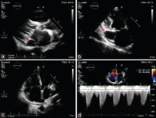
          Herein, we report the case of a 44-year-old female with end-stage renal disease on hemodialysis. She was admitted to our hospital to evaluate if she was eligible for a kidney transplant. Transthoracic echocardiography showed a markedly dilated coronary sinus and an unexpected finding of increased right ventriculoatrial gradient. A saline contrast echocardiography to confirm the presence of persistent left superior vena cava (PLSVC) was not performed because of arteriovenous fistula (FAV) for hemodialysis on the left forearm. Therefore, computed tomography angiography was performed, and it showed the PLSVC. We also proceeded with a transesophageal echocardiography which showed an atrial septal defect (ASD) of the sinus venous type hemodynamically significant. In this case, we identified a rare association of PLSVC with a ASD; therefore, there is a right ventricular volume overload because of the ASD hemodynamically significant and high flow FAV leading to a condition of a seeming pulmonary hypertension.

          Related collections

          Most cited references11

          • Record: found
          • Abstract: found
          • Article: found
          Is Open Access

          Persistent left superior vena cava: a case report and review of literature

          Persistent left superior vena cava is rare but important congenital vascular anomaly. It results when the left superior cardinal vein caudal to the innominate vein fails to regress. It is most commonly observed in isolation but can be associated with other cardiovascular abnormalities including atrial septal defect, bicuspid aortic valve, coarctation of aorta, coronary sinus ostial atresia, and cor triatriatum. The presence of PLSVC can render access to the right side of heart challenging via the left subclavian approach, which is a common site of access utilized when placing pacemakers and Swan-Ganz catheters. Incidental notation of a dilated coronary sinus on echocardiography should raise the suspicion of PLSVC. The diagnosis should be confirmed by saline contrast echocardiography.
            Bookmark
            • Record: found
            • Abstract: found
            • Article: not found

            Persistent left superior vena cava (PLSVC) with anomalous left hepatic vein drainage into the right atrium: role of imaging and clinical relevance.

            Persistent left superior vena cava (PLSVC) is a very rare and yet the most commonly described thoracic venous anomaly in medical literature. It has a 10-fold higher incidence with congenital heart disease. PLSVC often becomes apparent when an unknown PLSVC is incidentally discovered during central venous line placement, intracardiac electrode/pacemaker placement or cardiopulmonary bypass, where it may cause technical difficulties and life-threatening complications. PLSVC is also associated with disturbances of cardiac impulse formation and conduction including varying degrees of heart blocks, supraventricular arrhythmias and Wolff Parkinson White syndrome. We describe the case of an 86-year-old male with a history of coronary artery disease and chronic atrial fibrillation who presented with worsening dyspnea and syncopal episodes. An ECG was consistent with complete heart block. During lead placement for the pacemaker, a left subclavian approach was unsuccessful. A left venogram was performed through the brachial vein that demonstrated a left superior vena cava. The diagnosis was confirmed with echocardiography using a bubble study and also a chest CT. The anatomy was unique as there was anomalous left hepatic vein drainage into the right atrium. The case provides insight into the diagnostic modalities and clinical considerations of this unusual thoracic venous anomaly.
              Bookmark
              • Record: found
              • Abstract: found
              • Article: not found

              Transesophageal echocardiography is superior to transthoracic echocardiography in the diagnosis of sinus venosus atrial septal defect

              The purpose of this study was to compare transthoracic and transesophageal echocardiography in the diagnosis of various types of atrial septal defects. Forty-one adult patients with the clinical diagnosis of atrial septal defect were studied by transthoracic and transesophageal echocardiography (30 women, 11 men; 18 to 81 years of age). Transthoracic echocardiography demonstrated the atrial septal defect in 33 patients (secundum type in 28, primum type in 3 and sinus venosus type in 2). Transesophageal echocardiography demonstrated the defect in all 41 patients. Thus, in 8 (20%) of 41 patients the atrial septal defect was demonstrated by transesophageal and not by transthoracic echocardiography. Six of the eight had a sinus venosus type atrial septal defect; the other two patients had a secundum atrial septal defect (one of these two had a technically poor transthoracic echocardiogram and the other had a small atrial septal defect). Transthoracic echocardiography, therefore, failed to demonstrate the sinus venosus defect in six (75%) of eight patients. An anomalous venous connection associated with the sinus venosus defect was visualized by transesophageal echocardiography in seven of the eight patients but was not seen on transthoracic echocardiography in any patient. Sinus venosus type atrial septal defects are frequently not visualized in adults by conventional transthoracic echocardiography. Transesophageal echocardiography is recommended when an atrial septal defect is clinically suspected but cannot be visualized by transthoracic echocardiography.
                Bookmark

                Author and article information

                Journal
                J Cardiovasc Echogr
                J Cardiovasc Echogr
                JCE
                Journal of Cardiovascular Echography
                Medknow Publications & Media Pvt Ltd (India )
                2211-4122
                2347-193X
                Oct-Dec 2017
                : 27
                : 4
                : 149-152
                Affiliations
                [1]Cardiology Unit, University Hospital Policlinico, Bari, Italy
                Author notes
                Address for correspondence: Dr. Paolo Colonna, Cardiology Unit, University Hospital Policlinico, Piazza G. Cesare, 70124 Bari, Italy. E-mail: colonna@ 123456tiscali.it
                Article
                JCE-27-149
                10.4103/jcecho.jcecho_34_17
                5672689
                3c7fb07b-bdc3-4153-82c1-ada65802c5bd
                Copyright: © 2017 Journal of Cardiovascular Echography

                This is an open access article distributed under the terms of the Creative Commons Attribution-NonCommercial-ShareAlike 3.0 License, which allows others to remix, tweak, and build upon the work non-commercially, as long as the author is credited and the new creations are licensed under the identical terms.

                History
                Categories
                Case Report

                dilated coronary sinus,high-flow arteriovenous fistula,persistent left superior vena cava,sinus venous atrial septal defect

                Comments

                Comment on this article