19
views
0
recommends
+1 Recommend
0 collections
    0
    shares
      • Record: found
      • Abstract: found
      • Article: found
      Is Open Access

      Abortigenic but Not Neurotropic Equine Herpes Virus 1 Modulates the Interferon Antiviral Defense

      research-article

      Read this article at

      Bookmark
          There is no author summary for this article yet. Authors can add summaries to their articles on ScienceOpen to make them more accessible to a non-specialist audience.

          Abstract

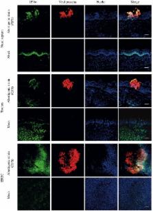
          Equine herpesvirus 1 (EHV1) is considered as a major pathogen of Equidae, causing symptoms from mild respiratory disease to late-term abortion and neurological disorders. Different EHV1 strains circulating in the field have been characterized to be of abortigenic or neurovirulent phenotype. Both variants replicate in a plaque-wise manner in the epithelium of the upper respiratory tract (URT), where the abortigenic strains induce more prominent viral plaques, compared to the neurovirulent strains. Considering the differences in replication at the URT, we hypothesized that abortigenic strains may show an increased ability to modulate the type I IFN secretion/signaling pathway, compared to strains that display the neurovirulent phenotype. Here, we analyze IFN levels induced by abortigenic and neurovirulent EHV1 using primary respiratory epithelial cells (EREC) and respiratory mucosa ex vivo explants. Similar levels of IFNα (~70 U/ml) were detected in explants inoculated with both types of EHV1 strains from 48 to 72 hpi. Second, EREC and mucosa explants were treated with recombinant equine IFNα (rEqIFNα) or Ruxolitinib (Rux), an IFN signaling inhibitor, prior to and during inoculation with abortigenic or neurovirulent EHV1. Replication of both EHV1 variants was suppressed by rEqIFNα. Further, addition of Rux increased replication in a concentration-dependent manner, indicating an IFN-susceptibility for both variants. However, in two out of three horses, at a physiological concentration of 100 U/ml of rEqIFNα, an increase in abortigenic EHV1 replication was observed compared to 10 U/ml of rEqIFNα, which was not observed for the neurovirulent strains. Moreover, in the presence of Rux, the plaque size of the abortigenic variants remained unaltered, whereas the typically smaller viral plaques induced by the neurovirulent variants became larger. Overall, our results demonstrate the importance of IFNα in the control of EHV1 replication in the URT for both abortigenic and neurovirulent variants. In addition, our findings support the speculation that abortigenic variants of EHV1 may have developed anti-IFN mechanisms that appear to be absent or less pronounced in neurovirulent EHV1 strains.

          Related collections

          Most cited references67

          • Record: found
          • Abstract: found
          • Article: not found

          The airway epithelium: soldier in the fight against respiratory viruses.

          The airway epithelium acts as a frontline defense against respiratory viruses, not only as a physical barrier and through the mucociliary apparatus but also through its immunological functions. It initiates multiple innate and adaptive immune mechanisms which are crucial for efficient antiviral responses. The interaction between respiratory viruses and airway epithelial cells results in production of antiviral substances, including type I and III interferons, lactoferrin, β-defensins, and nitric oxide, and also in production of cytokines and chemokines, which recruit inflammatory cells and influence adaptive immunity. These defense mechanisms usually result in rapid virus clearance. However, respiratory viruses elaborate strategies to evade antiviral mechanisms and immune responses. They may disrupt epithelial integrity through cytotoxic effects, increasing paracellular permeability and damaging epithelial repair mechanisms. In addition, they can interfere with immune responses by blocking interferon pathways and by subverting protective inflammatory responses toward detrimental ones. Finally, by inducing overt mucus secretion and mucostasis and by paving the way for bacterial infections, they favor lung damage and further impair host antiviral mechanisms.
            Bookmark
            • Record: found
            • Abstract: found
            • Article: not found

            Interferons and viruses: an evolutionary arms race of molecular interactions.

            Over half a century has passed since interferons (IFNs) were discovered and shown to inhibit virus infection in cultured cells. Since then, researchers have steadily brought to light the molecular details of IFN signaling, catalogued their pleiotropic effects on cells, and harnessed their therapeutic potential for a variety of maladies. While advances have been plentiful, several fundamental questions have yet to be answered and much complexity remains to be unraveled. We explore the current knowledge surrounding four main questions: are type I IFN subtypes differentially produced in response to distinct pathogens? How are IFN subtypes distinguished by cells? What are the mechanisms and consequences of viral antagonism? Lastly, how can the IFN response be harnessed to improve vaccine efficacy?
              Bookmark
              • Record: found
              • Abstract: found
              • Article: not found

              The interferons and their receptors—distribution and regulation

              The interferons (IFNs) were originally described over 50 years ago, identified by their ability to confer viral resistance to cells. We now know that they are much more than just anti‐viral cytokines collectively having roles in both innate and adaptive immune responses, in tumor surveillance and defense, and modulation of immune cell function. Three types of IFN have now been described, simply referred to as type I, II and III. Distinguishable by the unique receptors that they rely on for signal transduction, the three types of IFN have specific and varied roles in the maintenance of human health and defense against pathogens. In mounting an IFN‐mediated immune response, the human body has developed the ability to regulate IFN‐mediated signal transduction. Like all cytokines, the ability of a cell to respond to IFN is completely dependent on the presence of its cognate receptor on the surface of the target cell. Thus, one of the major mechanisms used by the human body to regulate the strength and duration of the IFN response is through regulation of receptor levels, thereby altering the cytokine‐specific responsiveness of the target cell. This review will discuss the receptor system utilized by the type I IFNs and compare it with that of the type II and III IFNs, which also regulate immune responses through controlling receptor level on the cell surface.
                Bookmark

                Author and article information

                Contributors
                Journal
                Front Cell Infect Microbiol
                Front Cell Infect Microbiol
                Front. Cell. Infect. Microbiol.
                Frontiers in Cellular and Infection Microbiology
                Frontiers Media S.A.
                2235-2988
                12 September 2018
                2018
                : 8
                : 312
                Affiliations
                [1] 1Department of Virology, Immunology and Parasitology, Faculty of Veterinary Medicine, Ghent University , Merelbeke, Belgium
                [2] 2Lewis Thomas Laboratory, Department of Molecular Biology, Princeton Neuroscience Institute, Princeton University , Princeton, NJ, United States
                [3] 3Department of Pathobiology and Diagnostic Investigation, College of Veterinary Medicine, Michigan State University , East Lansing, MI, United States
                Author notes

                Edited by: Rachel L. Roper, The Brody School of Medicine at East Carolina University, United States

                Reviewed by: Lara Maxwell, Oklahoma State University, United States; Abel Viejo-Borbolla, Hannover Medical School, Germany

                *Correspondence: Hans J. Nauwynck hans.nauwynck@ 123456ugent.be

                This article was submitted to Virus and Host, a section of the journal Frontiers in Cellular and Infection Microbiology

                Article
                10.3389/fcimb.2018.00312
                6144955
                30258819
                3c951a3e-8dcd-4bfd-a665-c5731321509a
                Copyright © 2018 Poelaert, Van Cleemput, Laval, Favoreel, Soboll Hussey, Maes and Nauwynck.

                This is an open-access article distributed under the terms of the Creative Commons Attribution License (CC BY). The use, distribution or reproduction in other forums is permitted, provided the original author(s) and the copyright owner(s) are credited and that the original publication in this journal is cited, in accordance with accepted academic practice. No use, distribution or reproduction is permitted which does not comply with these terms.

                History
                : 05 March 2018
                : 15 August 2018
                Page count
                Figures: 7, Tables: 0, Equations: 0, References: 84, Pages: 18, Words: 12548
                Funding
                Funded by: Agentschap voor Innovatie door Wetenschap en Technologie 10.13039/501100003132
                Award ID: 141627
                Funded by: Fonds Wetenschappelijk Onderzoek 10.13039/501100003130
                Award ID: 11Y5415N
                Categories
                Cellular and Infection Microbiology
                Original Research

                Infectious disease & Microbiology
                equine herpesvirus 1,upper respiratory tract,innate immunity,type i interferon,phenotype

                Comments

                Comment on this article