30
views
0
recommends
+1 Recommend
0 collections
    0
    shares
      • Record: found
      • Abstract: found
      • Article: not found

      GILT is a critical host factor for Listeria monocytogenes infection

      research-article
      1 , 2 , 1 , 1 , 2
      Nature

      Read this article at

      ScienceOpenPublisherPMC
      Bookmark
          There is no author summary for this article yet. Authors can add summaries to their articles on ScienceOpen to make them more accessible to a non-specialist audience.

          Abstract

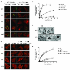
          Listeria monocytogenes is a gram positive, intracellular, food-borne pathogen that can cause severe illness in humans and animals. Upon infection, it is actively phagocytosed by macrophages 1. It then escapes from the phagosome, replicates in the cytosol, and subsequently spreads from cell to cell by a non-lytic mechanism driven by actin polymerization 2. Penetration of the phagosomal membrane is initiated by the secreted hemolysin listeriolysin O (LLO), which is essential for vacuolar escape in vitro and for virulence in animal models of infection 3. Reduction is required to activate the lytic activity of LLO in vitro 46, and we show here that reduction by the enzyme Gamma-interferon Inducible Lysosomal Thiolreductase (GILT) is responsible for the activation of LLO in vivo. GILT is a soluble thiol reductase expressed constitutively within the lysosomes of antigen presenting cells 7, 8, and it accumulates in macrophage phagosomes as they mature into phagolysosomes 9. The enzyme is delivered by a mannose-6-phosphate receptor-dependent mechanism to the endocytic pathway, where N- and C-terminal pro-peptides are cleaved to generate a 30 kDa mature enzyme 7, 8, 10. The active site of GILT contains two cysteine residues in a CXXC motif that catalyzes the reduction of disulfide bonds 7, 8. Mice lacking GILT are deficient in generating MHC class II-restricted CD4 + T cell responses to protein antigens that contain disulfide bonds 11, 12. Here we show that these mice are resistant to L. monocytogenes infection. Replication of the organism in GILT-negative macrophages, or macrophages expressing an enzymatically inactive GILT mutant, is impaired because of delayed escape from the phagosome. GILT activates LLO within the phagosome by the classical thiol reductase mechanism shared by members of the thioredoxin family. In addition, purified GILT activates recombinant LLO, facilitating membrane permeabilization and red blood cell lysis. The data show GILT is a critical host factor that facilitates L. monocytogenes infection.

          Related collections

          Most cited references27

          • Record: found
          • Abstract: found
          • Article: not found

          Actin filaments and the growth, movement, and spread of the intracellular bacterial parasite, Listeria monocytogenes

          Listeria monocytogenes was used as a model intracellular parasite to study stages in the entry, growth, movement, and spread of bacteria in a macrophage cell line. The first step in infection is phagocytosis of the Listeria, followed by the dissolution of the membrane surrounding the phagosome presumably mediated by hemolysin secreted by Listeria as nonhemolytic mutants remain in intact vacuoles. Within 2 h after infection, each now cytoplasmic Listeria becomes encapsulated by actin filaments, identified as such by decoration of the actin filaments with subfragment 1 of myosin. These filaments are very short. The Listeria grow and divide and the actin filaments rearrange to form a long tail (often 5 microns in length) extending from only one end of the bacterium, a "comet's tail," in which the actin filaments appear randomly oriented. The Listeria "comet" moves to the cell surface with its tail oriented towards the cell center and becomes incorporated into a cell extension with the Listeria at the tip of the process and its tail trailing into the cytoplasm behind it. This extension contacts a neighboring macrophage that phagocytoses the extension of the first macrophage. Thus, within the cytoplasm of the second macrophage is a Listeria with its actin tail surrounded by a membrane that in turn is surrounded by the phagosome membrane of the new host. Both these membranes are then solubilized by the Listeria and the cycle is repeated. Thus, once inside a host cell, the infecting Listeria and their progeny can spread from cell to cell by remaining intracellular and thus bypass the humoral immune system of the organism. To establish if actin filaments are essential for the spread of Listeria from cell to cell, we treated infected macrophages with cytochalasin D. The Listeria not only failed to spread, but most were found deep within the cytoplasm, rather than near the periphery of the cell. Thin sections revealed that the net of actin filaments is not formed nor is a "comet" tail produced.
            Bookmark
            • Record: found
            • Abstract: found
            • Article: not found

            The Phagosome Proteome

            Phagosomes are key organelles for the innate ability of macrophages to participate in tissue remodeling, clear apoptotic cells, and restrict the spread of intracellular pathogens. To understand the functions of phagosomes, we initiated the systematic identification of their proteins. Using a proteomic approach, we identified >140 proteins associated with latex bead–containing phagosomes. Among these were hydrolases, proton pump ATPase subunits, and proteins of the fusion machinery, validating our approach. A series of unexpected proteins not previously described along the endocytic/phagocytic pathways were also identified, including the apoptotic proteins galectin3, Alix, and TRAIL, the anti-apoptotic protein 14-3-3, the lipid raft-enriched flotillin-1, the anti-microbial molecule lactadherin, and the small GTPase rab14. In addition, 24 spots from which the peptide masses could not be matched to entries in any database potentially represent new phagosomal proteins. The elaboration of a two-dimensional gel database of >160 identified spots allowed us to analyze how phagosome composition is modulated during phagolysosome biogenesis. Remarkably, during this process, hydrolases are not delivered in bulk to phagosomes, but are instead acquired sequentially. The systematic characterization of phagosome proteins provided new insights into phagosome functions and the protein or groups of proteins involved in and regulating these functions.
              Bookmark
              • Record: found
              • Abstract: found
              • Article: not found

              Genome-wide RNAi screen for host factors required for intracellular bacterial infection.

              Most studies of host-pathogen interactions have focused on pathogen-specific virulence determinants. Here, we report a genome-wide RNA interference screen to identify host factors required for intracellular bacterial pathogenesis. Using Drosophila cells and the cytosolic pathogen Listeria monocytogenes, we identified 305 double-stranded RNAs targeting a wide range of cellular functions that altered L. monocytogenes infection. Comparison to a similar screen with Mycobacterium fortuitum, a vacuolar pathogen, identified host factors that may play a general role in intracellular pathogenesis and factors that specifically affect access to the cytosol by L. monocytogenes.
                Bookmark

                Author and article information

                Journal
                0410462
                6011
                Nature
                Nature
                0028-0836
                1476-4687
                24 April 2009
                24 September 2008
                30 October 2008
                10 November 2009
                : 455
                : 7217
                : 1244-1247
                Affiliations
                [1 ]Department of Immunobiology, Yale University School of Medicine, 300 Cedar St., New Haven, Connecticut 06250-8011
                [2 ]Howard Hughes Medical Institute, Yale University School of Medicine, 300 Cedar St., New Haven, Connecticut 06250-8011
                Author notes
                Corresponding Author Info: Peter Cresswell, Ph.D., Department of Immunobiology, Howard Hughes Medical Institute, Yale University School of Medicine, 300 Cedar Street, P.O. Box 208011, New Haven, CT 06520-8011, Ph: 203-785-5176, Fax: 203-785-4461, peter.cresswell@ 123456yale.edu
                [3]

                Present Address: Max F. Perutz Laboratories GmbH, Rennweg 95a, 1030 Wien 3, Landstraβe, Wiene, Austria

                Author Contributions

                RS performed experiments, AJ assisted with the in vivo infection and RS and PC wrote the paper.

                Reprints and permissions information is available at www.nature.com/reprints.

                Article
                nihpa67290
                10.1038/nature07344
                2775488
                18815593
                3cfa3a61-b056-4ee3-812b-284f6b22e1e6
                History
                Funding
                Funded by: National Institute of Allergy and Infectious Diseases Extramural Activities : NIAID
                Award ID: R37 AI023081-24 ||AI
                Categories
                Article

                Uncategorized
                Uncategorized

                Comments

                Comment on this article