59
views
0
recommends
+1 Recommend
0 collections
    0
    shares
      • Record: found
      • Abstract: found
      • Article: found
      Is Open Access

      Electrical impedance tomography measured at two thoracic levels can visualize the ventilation distribution changes at the bedside during a decremental positive end-expiratory lung pressure trial

      research-article

      Read this article at

      Bookmark
          There is no author summary for this article yet. Authors can add summaries to their articles on ScienceOpen to make them more accessible to a non-specialist audience.

          Abstract

          Introduction

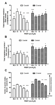
          Computed tomography of the lung has shown that ventilation shifts from dependent to nondependent lung regions. In this study, we investigated whether, at the bedside, electrical impedance tomography (EIT) at the cranial and caudal thoracic levels can be used to visualize changes in ventilation distribution during a decremental positive end-expiratory pressure (PEEP) trial and the relation of these changes to global compliance in mechanically ventilated patients.

          Methods

          Ventilation distribution was calculated on the basis of EIT results from 12 mechanically ventilated patients after cardiac surgery at a cardiothoracic ICU. Measurements were taken at four PEEP levels (15, 10, 5 and 0 cm H 2O) at both the cranial and caudal lung levels, which were divided into four ventral-to-dorsal regions. Regional compliance was calculated using impedance and driving pressure data.

          Results

          We found that tidal impedance variation divided by tidal volume significantly decreased on caudal EIT slices, whereas this measurement increased on the cranial EIT slices. The dorsal-to-ventral impedance distribution, expressed according to the center of gravity index, decreased during the decremental PEEP trial at both EIT levels. Optimal regional compliance differed at different PEEP levels: 10 and 5 cm H 2O at the cranial level and 15 and 10 cm H 2O at the caudal level for the dependent and nondependent lung regions, respectively.

          Conclusions

          At the bedside, EIT measured at two thoracic levels showed different behavior between the caudal and cranial lung levels during a decremental PEEP trial. These results indicate that there is probably no single optimal PEEP level for all lung regions.

          Related collections

          Most cited references22

          • Record: found
          • Abstract: not found
          • Article: not found

          What has computed tomography taught us about the acute respiratory distress syndrome?

            Bookmark
            • Record: found
            • Abstract: found
            • Article: not found

            Imbalances in regional lung ventilation: a validation study on electrical impedance tomography.

            Imbalances in regional lung ventilation, with gravity-dependent collapse and overdistention of nondependent zones, are likely associated to ventilator-induced lung injury. Electric impedance tomography is a new imaging technique that is potentially capable of monitoring those imbalances. The aim of this study was to validate electrical impedance tomography measurements of ventilation distribution, by comparison with dynamic computerized tomography in a heterogeneous population of critically ill patients under mechanical ventilation. Multiple scans with both devices were collected during slow-inflation breaths. Six repeated breaths were monitored by impedance tomography, showing acceptable reproducibility. We observed acceptable agreement between both technologies in detecting right-left ventilation imbalances (bias = 0% and limits of agreement = -10 to +10%). Relative distribution of ventilation into regions or layers representing one-fourth of the thoracic section could also be assessed with good precision. Depending on electrode positioning, impedance tomography slightly overestimated ventilation imbalances along gravitational axis. Ventilation was gravitationally dependent in all patients, with some transient blockages in dependent regions synchronously detected by both scanning techniques. Among variables derived from computerized tomography, changes in absolute air content best explained the integral of impedance changes inside regions of interest (r(2) > or = 0.92). Impedance tomography can reliably assess ventilation distribution during mechanical ventilation.
              Bookmark
              • Record: found
              • Abstract: found
              • Article: not found

              Lung opening and closing during ventilation of acute respiratory distress syndrome.

              The effects of high positive end-expiratory pressure (PEEP) strictly depend on lung recruitability, which varies widely during acute respiratory distress syndrome (ARDS). Unfortunately, increasing PEEP may lead to opposing effects on two main factors potentially worsening the lung injury, that is, alveolar strain and intratidal opening and closing, being detrimental (increasing the former) or beneficial (decreasing the latter). To investigate how lung recruitability influences alveolar strain and intratidal opening and closing after the application of high PEEP. We analyzed data from a database of 68 patients with acute lung injury or ARDS who underwent whole-lung computed tomography at 5, 15, and 45 cm H(2)O airway pressure. End-inspiratory nonaerated lung tissue was estimated from computed tomography pressure-volume curves. Alveolar strain and opening and closing lung tissue were computed at 5 and 15 cm H(2)O PEEP. In patients with a higher percentage of potentially recruitable lung, the increase in PEEP markedly reduced opening and closing lung tissue (P < 0.001), whereas no differences were observed in patients with a lower percentage of potentially recruitable lung. In contrast, alveolar strain similarly increased in the two groups (P = 0.89). Opening and closing lung tissue was distributed mainly in the dependent and hilar lung regions, and it appeared to be an independent risk factor for death (odds ratio, 1.10 for each 10-g increase). In ARDS, especially in patients with higher lung recruitability, the beneficial impact of reducing intratidal alveolar opening and closing by increasing PEEP prevails over the effects of increasing alveolar strain.
                Bookmark

                Author and article information

                Journal
                Crit Care
                Crit Care
                Critical Care
                BioMed Central
                1364-8535
                1466-609X
                2011
                11 August 2011
                : 15
                : 4
                : R193
                Affiliations
                [1 ]Department of Intensive Care Medicine, Erasmus MC, 's-Gravendijkwal 230, NL-3015GE Rotterdam, The Netherlands
                [2 ]Department of Anesthesiology, Erasmus MC, 's-Gravendijkwal 230, NL-3015GE Rotterdam, The Netherlands
                Article
                cc10354
                10.1186/cc10354
                3387635
                21834953
                3d3765cc-22e3-4a8a-a9b8-b174ee115753
                Copyright ©2011 Bikker et al.; licensee BioMed Central Ltd.

                This is an open access article distributed under the terms of the Creative Commons Attribution License ( http://creativecommons.org/licenses/by/2.0), which permits unrestricted use, distribution, and reproduction in any medium, provided the original work is properly cited.

                History
                : 1 October 2010
                : 12 July 2011
                : 11 August 2011
                Categories
                Research

                Emergency medicine & Trauma
                electric impedance,mechanical ventilation,positive-pressure respiration,humans,atelectasis,critical care

                Comments

                Comment on this article