29
views
0
recommends
+1 Recommend
0 collections
    0
    shares
      • Record: found
      • Abstract: found
      • Article: found
      Is Open Access

      Cyp19a1 (Aromatase) Expression in the Xenopus Brain at Different Developmental Stages

      research-article
      ,
      Journal of Neuroendocrinology
      BlackWell Publishing Ltd
      Aromatase, Cyp19a1, radial glia, Xenopus, Brain

      Read this article at

      Bookmark
          There is no author summary for this article yet. Authors can add summaries to their articles on ScienceOpen to make them more accessible to a non-specialist audience.

          Abstract

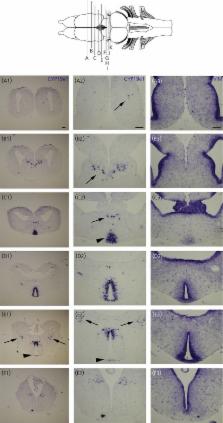
          Cytochrome P450 aromatase (P450arom; aromatase) is a microsomal enzyme involved in the production of endogeneous sex steroids by converting testosterone into oestradiol. Aromatase is the product of the cyp19a1 gene and plays a crucial role in the sexual differentiation of the brain and in the regulation of reproductive functions. In the brain of mammals and birds, expression of cyp19a1 has been demonstrated in neuronal populations of the telencephalon and diencephalon. By contrast, a wealth of evidence established that, in teleost fishes, aromatase expression in the brain is restricted to radial glial cells. The present study investigated the precise neuroanatomical distribution of cyp19a1 mRNA during brain development in Xenopus laevis (late embryonic to juvenile stages). For this purpose, we used in situ hybridisation alone or combined with the detection of a proliferative (proliferating cell nuclear antigen), glial (brain lipid binding protein, Vimentin) or neuronal (acetylated tubulin; HuC/D; NeuroβTubulin) markers. We provide evidence that cyp19a1 expression in the brain is initiated from the very early larval stage and remains strongly detected until the juvenile and adult stages. At all stages analysed, we found the highest expression of cyp19a1 in the preoptic area and the hypothalamus compared to the rest of the brain. In these two brain regions, cyp19a1-positive cells were never detected in the ventricular layers. Indeed, no co-labelling could be observed with radial glial (brain lipid binding protein, Vimentin) or dividing progenitors (proliferating cell nuclear antigen) markers. By contrast, cyp19a1-positive cells perfectly matched with the distribution of post-mitotic neurones as shown by the use of specific markers (HuC/D, acetylated tubulin and NeuroβTubulin). These data suggest that, similar to that found in other tetrapods, aromatase in the brain of amphibians is found in post-mitotic neurones and not in radial glia as reported in teleosts.

          Related collections

          Most cited references79

          • Record: found
          • Abstract: found
          • Article: not found

          Aromatase--a brief overview.

          There is growing awareness that androgens and estrogens have general metabolic roles that are not directly involved in reproductive processes. These include actions on vascular function, lipid and carbohydrate metabolism, as well as bone mineralization and epiphyseal closure in both sexes. In postmenopausal women, as in men, estrogen is no longer solely an endocrine factor but instead is produced in a number of extragonadal sites and acts locally at these sites in a paracrine and intracrine fashion. These sites include breast, bone, vasculature, and brain. Within these sites, aromatase action can generate high levels of estradiol locally without significantly affecting circulating levels. Circulating C19 steroid precursors are essential substrates for extragonadal estrogen synthesis. The levels of these androgenic precursors decline markedly with advancing age in women, possible from the mid-to-late reproductive years. This may be a fundamental reason why women are at increased risk for bone mineral loss and fracture, and possibly decline of cognitive function, compared with men. Aromatase expression in these various sites is under the control of tissue-specific promotors regulated by different cohorts of transcription factors. Thus in principle, it should be possible to develop selective aromatase modulators (SAMs) that block aromatase expression, for example, in breast, but allow unimpaired estrogen synthesis in other tissues such as bone.
            Bookmark
            • Record: found
            • Abstract: found
            • Article: not found

            Estrogen masculinizes neural pathways and sex-specific behaviors.

            Sex hormones are essential for neural circuit development and sex-specific behaviors. Male behaviors require both testosterone and estrogen, but it is unclear how the two hormonal pathways intersect. Circulating testosterone activates the androgen receptor (AR) and is also converted into estrogen in the brain via aromatase. We demonstrate extensive sexual dimorphism in the number and projections of aromatase-expressing neurons. The masculinization of these cells is independent of AR but can be induced in females by either testosterone or estrogen, indicating a role for aromatase in sexual differentiation of these neurons. We provide evidence suggesting that aromatase is also important in activating male-specific aggression and urine marking because these behaviors can be elicited by testosterone in males mutant for AR and in females subjected to neonatal estrogen exposure. Our results suggest that aromatization of testosterone into estrogen is important for the development and activation of neural circuits that control male territorial behaviors.
              Bookmark
              • Record: found
              • Abstract: found
              • Article: not found

              Considering the evolution of regeneration in the central nervous system.

              For many years the mammalian CNS has been seen as an organ that is unable to regenerate. However, it was also long known that lower vertebrate species are capable of impressive regeneration of CNS structures. How did this situation arise through evolution? Increasing cellular and molecular understanding of regeneration in different animal species coupled with studies of adult neurogenesis in mammals is providing a basis for addressing this question. Here we compare CNS regeneration among vertebrates and speculate on how this ability may have emerged or been restricted.
                Bookmark

                Author and article information

                Journal
                J Neuroendocrinol
                J. Neuroendocrinol
                jne
                Journal of Neuroendocrinology
                BlackWell Publishing Ltd (Oxford, UK )
                0953-8194
                1365-2826
                April 2014
                03 April 2014
                : 26
                : 4
                : 226-236
                Affiliations
                Neuroendocrine Effects of Endocrine Disruptors, IRSET, INSERM U1085, SFR Biosit, Université de Rennes 1 Rennes, France
                Author notes
                Correspondence to: Pascal Coumailleau, IRSET, INSERM U1085, Université de Rennes1, Campus de Beaulieu, Bat. 13, Avenue du Général Leclerc, 35042 Rennes, France (e-mail: Pascal.coumailleau@ 123456univ-rennes1.fr )
                Article
                10.1111/jne-12142
                4238815
                24612124
                3eda9b19-502c-4f6d-99cf-7995b2538952
                © 2014 The Authors. Journal of Neuroendocrinology published by John Wiley & Sons Ltd on behalf of The British Society for Neuroendocrinology.

                This is an open access article under the terms of the Creative Commons Attribution License, which permits use, distribution and reproduction in any medium, provided the original work is properly cited.

                History
                : 20 November 2013
                : 17 February 2014
                : 22 February 2014
                Categories
                Original Articles

                Endocrinology & Diabetes
                aromatase,cyp19a1,radial glia,xenopus,brain
                Endocrinology & Diabetes
                aromatase, cyp19a1, radial glia, xenopus, brain

                Comments

                Comment on this article