10
views
0
recommends
+1 Recommend
0 collections
    0
    shares
      • Record: found
      • Abstract: found
      • Article: found
      Is Open Access

      Ursolic acid supplementation decreases markers of skeletal muscle damage during resistance training in resistance-trained men: a pilot study

      research-article

      Read this article at

      Bookmark
          There is no author summary for this article yet. Authors can add summaries to their articles on ScienceOpen to make them more accessible to a non-specialist audience.

          Abstract

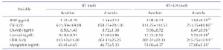
          Ursolic acid (UA) supplementation was previously shown to improve skeletal muscle function in resistance-trained men. This study aimed to determine, using the same experimental paradigm, whether UA also has beneficial effects on exercise-induced skeletal muscle damage markers including the levels of cortisol, B-type natriuretic peptide (BNP), myoglobin, creatine kinase (CK), creatine kinase-myocardial band (CK-MB), and lactate dehydrogenase (LDH) in resistance-trained men. Sixteen healthy participants were randomly assigned to resistance training (RT) or RT+UA groups (n=8 per group). Participants were trained according to the RT program (60~80% of 1 repetition, 6 times/week), and the UA group was additionally given UA supplementation (450 mg/day) for 8 weeks. Blood samples were obtained before and after intervention, and cortisol, BNP, myoglobin, CK, CK-MB, and LDH levels were analyzed. Subjects who underwent RT alone showed no significant change in body composition and markers of skeletal muscle damage, whereas RT+UA group showed slightly decreased body weight and body fat percentage and slightly increased lean body mass, but without statistical significance. In addition, UA supplementation significantly decreased the BNP, CK, CK-MB, and LDH levels (p<0.05). In conclusion, UA supplementation alleviates increased skeletal muscle damage markers after RT. This finding provides evidence for a potential new therapy for resistance-trained men.

          Related collections

          Most cited references37

          • Record: found
          • Abstract: found
          • Article: not found

          mRNA expression signatures of human skeletal muscle atrophy identify a natural compound that increases muscle mass.

          Skeletal muscle atrophy is a common and debilitating condition that lacks a pharmacologic therapy. To develop a potential therapy, we identified 63 mRNAs that were regulated by fasting in both human and mouse muscle, and 29 mRNAs that were regulated by both fasting and spinal cord injury in human muscle. We used these two unbiased mRNA expression signatures of muscle atrophy to query the Connectivity Map, which singled out ursolic acid as a compound whose signature was opposite to those of atrophy-inducing stresses. A natural compound enriched in apples, ursolic acid reduced muscle atrophy and stimulated muscle hypertrophy in mice. It did so by enhancing skeletal muscle insulin/IGF-I signaling and inhibiting atrophy-associated skeletal muscle mRNA expression. Importantly, ursolic acid's effects on muscle were accompanied by reductions in adiposity, fasting blood glucose, and plasma cholesterol and triglycerides. These findings identify a potential therapy for muscle atrophy and perhaps other metabolic diseases. Copyright © 2011 Elsevier Inc. All rights reserved.
            Bookmark
            • Record: found
            • Abstract: found
            • Article: not found

            Muscle function after exercise-induced muscle damage and rapid adaptation.

            This brief review focuses on the time course of changes in muscle function and other correlates of muscle damage following maximal effort eccentric actions of the forearm flexor muscles. Data on 109 subjects are presented to describe an accurate time course of these changes and attempt to establish relationships among the measures. Peak soreness is experienced 2-3 d postexercise while peak swelling occurs 5 d postexercise. Maximal strength and the ability to fully flex the arm show the greatest decrements immediately after exercise with a linear restoration of these functions over the next 10 d. Blood creatine kinase (CK) levels increase precipitously at 2 d after exercise which is also the time when spontaneous muscle shortening is most pronounced. Whether the similarity in the time courses of some of these responses implies that they are caused by similar factors remains to be determined. Performance of one bout of eccentric exercise produces an adaptation such that the muscle is more resistant to damage from a subsequent bout of exercise. The length of the adaptation differs among the measures such that when the exercise regimens are separated by 6 wk, all measures show a reduction in response on the second, compared with the first, bout. After 10 wk, only CK and muscle shortening show a reduction in response. After 6 months only the CK response is reduced. A combination of cellular factors and neurological factors may be involved in the adaptation process.
              Bookmark
              • Record: found
              • Abstract: found
              • Article: not found

              Skeletal muscle hypertrophy following resistance training is accompanied by a fiber type-specific increase in satellite cell content in elderly men.

              We determined muscle fiber type-specific hypertrophy and changes in satellite cell (SC) content following a 12-week resistance training program in 13 healthy, elderly men (72 +/- 2 years). Leg strength and body composition (dual-energy X-ray absorptiometry and computed tomography) were assessed, and muscle biopsy samples were collected. Leg strength increased 25%-30% after training (p < .001). Leg lean mass and quadriceps cross-sectional area increased 6%-9% (p < .001). At baseline, mean fiber area and SC content were smaller in the Type II versus Type I muscle fibers (p < .01). Following training, Type II muscle fiber area increased from 5,438 +/- 319 to 6,982 +/- 503 microm(2) (p < .01). Type II muscle fiber SC content increased from 0.048 +/- 0.003 to 0.084 +/- 0.008 SCs per fiber (p < .001). No changes were observed in the Type I muscle fibers. In older adults, skeletal muscle tissue is still capable of inducing SC proliferation and differentiation, resulting in Type II muscle fiber hypertrophy.
                Bookmark

                Author and article information

                Journal
                Korean J Physiol Pharmacol
                Korean J. Physiol. Pharmacol
                KJPP
                The Korean Journal of Physiology & Pharmacology : Official Journal of the Korean Physiological Society and the Korean Society of Pharmacology
                The Korean Physiological Society and The Korean Society of Pharmacology
                1226-4512
                2093-3827
                November 2017
                30 October 2017
                : 21
                : 6
                : 651-656
                Affiliations
                [1 ]Department of Physical Education, College of Health, Social Welfare and Education, Tong Myong University, Busan 48520, Korea.
                [2 ]National Research Laboratory for Mitochondrial Signaling, Department of Physiology, Department of Health Sciences and Technology, BK 21 Plus Team, College of Medicine, Cardiovascular and Metabolic Disease Center, Inje University, Busan 47392, Korea.
                [3 ]School of Free Major, Tong Myong University, Busan 48520, Korea.
                [4 ]Department of Physical Education, Changwon National University, Changwon 51140, Korea.
                [5 ]Department of Kinesiology, Inha University, Incheon 22212, Korea.
                [6 ]Department of Sports Leisure, College of Kyungsang, Busan 47583, Korea.
                [7 ]Department of Sports Industry, Busan University of Foreign Studies, Busan 46234, Korea.
                [8 ]Department of Sports Science, College of Natural Science, Chonbuk National University, Jeonju 54896, Korea.
                Author notes
                Correspondence: Jin Han. phyhanj@ 123456inje.ac.kr

                #These authors contributed equally to this work.

                Article
                10.4196/kjpp.2017.21.6.651
                5709482
                3f0f8a95-8ead-4203-9d10-413fbd6a91dc
                Copyright © 2017 The Korean Physiological Society and The Korean Society of Pharmacology

                This is an Open Access article distributed under the terms of the Creative Commons Attribution Non-Commercial License ( http://creativecommons.org/licenses/by-nc/4.0) which permits unrestricted non-commercial use, distribution, and reproduction in any medium, provided the original work is properly cited.

                History
                : 20 June 2017
                : 24 August 2017
                : 07 September 2017
                Funding
                Funded by: National Research Foundation of Korea, CrossRef http://dx.doi.org/10.13039/501100003725;
                Award ID: 2015R1A2A1A13001900
                Funded by: Ministry of Education, CrossRef http://dx.doi.org/10.13039/501100002701;
                Award ID: 2010-0020224
                Categories
                Original Article

                resistance training,resistance-trained men,skeletal muscle damage markers,ursolic acid

                Comments

                Comment on this article