47
views
0
recommends
+1 Recommend
0 collections
    0
    shares
      • Record: found
      • Abstract: found
      • Article: found
      Is Open Access

      miR-93 Promotes Cell Proliferation in Gliomas through Activation of PI3K/Akt Signaling Pathway

      research-article

      Read this article at

      Bookmark
          There is no author summary for this article yet. Authors can add summaries to their articles on ScienceOpen to make them more accessible to a non-specialist audience.

          Abstract

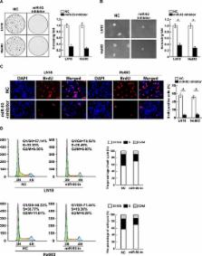
          The PI3K/Akt signaling pathway is frequently activated in various human cancer types and plays essential roles in development and progression of cancers. Multiple regulators, such as phosphatase and tensin homolog (PTEN) and PH domain leucine rich repeat protein phosphatases (PHLPP), have also found to be involved in suppression of the PI3K/Akt signaling pathway. However, how suppressive effects mediated by these regulators are concomitantly disrupted in cancers, which display constitutively activated PI3K/Akt signaling, remains puzzling. In the present study, we reported that the expression of miR-93 was markedly upregulated in glioma cell lines and clinical glioma tissues. Statistical analysis revealed that miR-93 levels significantly correlated with clinicopathologic grade and overall survival in gliomas. Furthermore, we found that overexpressing miR-93 promoted, but inhibition of miR-93 reduced, glioma cell proliferation and cell-cycle progression. We demonstrated that miR-93 activated PI3K/Akt signaling through directly suppressing PTEN, PHLPP2 and FOXO3 expression via targeting their 3′UTRs. Therefore, our results suggest that miR-93 might play an important role in glioma progression and uncover a novel mechanism for constitutive PI3K/Akt activation in gliomas.

          Related collections

          Most cited references45

          • Record: found
          • Abstract: found
          • Article: not found

          Targeting of the Bmi-1 oncogene/stem cell renewal factor by microRNA-128 inhibits glioma proliferation and self-renewal.

          MicroRNAs (miR) show characteristic expression signatures in various cancers and can profoundly affect cancer cell behavior. We carried out miR expression profiling of human glioblastoma specimens versus adjacent brain devoid of tumor. This revealed several significant alterations, including a pronounced reduction of miR-128 in tumor samples. miR-128 expression significantly reduced glioma cell proliferation in vitro and glioma xenograft growth in vivo. miR-128 caused a striking decrease in expression of the Bmi-1 oncogene, by direct regulation of the Bmi-1 mRNA 3'-untranslated region, through a single miR-128 binding site. In a panel of patient glioblastoma specimens, Bmi-1 expression was significantly up-regulated and miR-128 was down-regulated compared with normal brain. Bmi-1 functions in epigenetic silencing of certain genes through epigenetic chromatin modification. We found that miR-128 expression caused a decrease in histone methylation (H3K27me(3)) and Akt phosphorylation, and up-regulation of p21(CIP1) levels, consistent with Bmi-1 down-regulation. Bmi-1 has also been shown to promote stem cell self-renewal; therefore, we investigated the effects of miR-128 overexpression in human glioma neurosphere cultures, possessing features of glioma "stem-like" cells. This showed that miR-128 specifically blocked glioma self-renewal consistent with Bmi-1 down-regulation. This is the first example of specific regulation by a miR of a neural stem cell self-renewal factor, implicating miRs that may normally regulate brain development as important biological and therapeutic targets against the "stem cell-like" characteristics of glioma.
            Bookmark
            • Record: found
            • Abstract: not found
            • Article: not found

            AKT plays a central role in tumorigenesis.

              Bookmark
              • Record: found
              • Abstract: found
              • Article: not found

              Enumeration of the simian virus 40 early region elements necessary for human cell transformation.

              While it is clear that cancer arises from the accumulation of genetic mutations that endow the malignant cell with the properties of uncontrolled growth and proliferation, the precise combinations of mutations that program human tumor cell growth remain unknown. The study of the transforming proteins derived from DNA tumor viruses in experimental models of transformation has provided fundamental insights into the process of cell transformation. We recently reported that coexpression of the simian virus 40 (SV40) early region (ER), the gene encoding the telomerase catalytic subunit (hTERT), and an oncogenic allele of the H-ras gene in normal human fibroblast, kidney epithelial, and mammary epithelial cells converted these cells to a tumorigenic state. Here we show that the SV40 ER contributes to tumorigenic transformation in the presence of hTERT and oncogenic H-ras by perturbing three intracellular pathways through the actions of the SV40 large T antigen (LT) and the SV40 small t antigen (ST). LT simultaneously disables the retinoblastoma (pRB) and p53 tumor suppressor pathways; however, complete transformation of human cells requires the additional perturbation of protein phosphatase 2A by ST. Expression of ST in this setting stimulates cell proliferation, permits anchorage-independent growth, and confers increased resistance to nutrient deprivation. Taken together, these observations define the elements of the SV40 ER required for the transformation of human cells and begin to delineate a set of intracellular pathways whose disruption, in aggregate, appears to be necessary to generate tumorigenic human cells.
                Bookmark

                Author and article information

                Journal
                Oncotarget
                Oncotarget
                ImpactJ
                Oncotarget
                Impact Journals LLC
                1949-2553
                10 April 2015
                13 March 2015
                : 6
                : 10
                : 8286-8299
                Affiliations
                1 Department of Pathophysiology, Guangzhou Medical University, Guangzhou 510182, China
                2 State Key Laboratory of Oncology in Southern China, Department of Experimental Research, Cancer Centre, Sun Yat-sen University, Guangzhou 510060, China
                3 Department of the Central Laboratory, The First Affiliated Hospital/School of Clinical Medicine of Guangdong Pharmaceutical University, Guangzhou 510080, China
                4 Laboratory of Surgery, First Affiliated Hospital, Sun Yat-sen University, Guangzhou 510080, China
                5 Department of Biochemistry, Zhongshan School of Medicine, Sun Yat-sen University, Guangzhou, Guangdong 510080, China
                Author notes
                Correspondence to: Libing Song, lb.song1@ 123456gmail.com
                Article
                10.18632/oncotarget.3221
                4480752
                25823655
                44674f43-ef51-40e3-9ca8-9c6b8f653d61
                Copyright: © 2015 Jiang et al.

                This is an open-access article distributed under the terms of the Creative Commons Attribution License, which permits unrestricted use, distribution, and reproduction in any medium, provided the original author and source are credited.

                History
                : 2 December 2014
                : 26 January 2015
                Categories
                Research Paper

                Oncology & Radiotherapy
                pi3k/akt,mir-93,gliomas
                Oncology & Radiotherapy
                pi3k/akt, mir-93, gliomas

                Comments

                Comment on this article