8
views
0
recommends
+1 Recommend
0 collections
    0
    shares
      • Record: found
      • Abstract: found
      • Article: found
      Is Open Access

      Predictive factors of difficulty in lower third molar extraction: A prospective cohort study

      research-article

      Read this article at

      Bookmark
          There is no author summary for this article yet. Authors can add summaries to their articles on ScienceOpen to make them more accessible to a non-specialist audience.

          Abstract

          Background

          Several publications have measured the difficulty of third molar removal, trying to establish the main risk factors, however several important preoperative and intraoperative variables are overlooked.

          Material and Methods

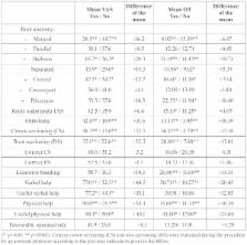
          A prospective cohort study comprising a total of 130 consecutive lower third molar extractions was performed. The outcome variables used to measure the difficulty of the extraction were operation time and a 100mm visual analogue scale filled by the surgeon at the end of the surgical procedure. The predictors were divided into 4 different groups (demographic, anatomic, radiographic and operative variables). A descriptive, bivariate and multivariate analysis of the data was performed.

          Results

          Patients’ weight, the presence of bulbous roots, the need to perform crown and root sectioning of the lower third molar and Pell and Gregory 123 classification significantly influenced both outcome variables ( p< 0.05).

          Conclusions

          Certain anatomical, radiological and operative variables appear to be important factors in the assessment of surgical difficulty in the extraction of lower third molars.

          Key words:Third molar, surgical extraction, surgical difficulty.

          Related collections

          Most cited references30

          • Record: found
          • Abstract: found
          • Article: not found

          The radiological prediction of inferior alveolar nerve injury during third molar surgery.

          The surgical removal of an impacted mandibular third molar may result in damage to the inferior alveolar nerve and may cause disabling anaesthesia of the lip; anaesthesia of the lower gingivae and anterior teeth may also result. Assessing the likelihood of injury depends to a great extent on preoperative radiographic examination. Seven radiological diagnostic signs have been mentioned in the literature; the reliability of these signs as predictors of likely nerve injury have been evaluated through retrospective and prospective surveys. Three signs were found to be significantly related to nerve injury and a further two were probably important clinically.
            Bookmark
            • Record: found
            • Abstract: found
            • Article: not found

            Factors predictive of difficulty of mandibular third molar surgery.

            Historically the difficulty of third molar surgery has been judged using radiologically assessed dental factors specifically tooth morphology and position. This study investigated additional factors that have a bearing on the difficulty of extraction. A prospective study undertaken by three clinical assistant grade surgeons who removed 354 single mandibular third molar teeth under day case anaesthesia over the 4-year period (1994-1998). Data relating to patient, dental and surgical variables were collected contemporaneously as the patients were treated. The difficulty of extraction was estimated by the surgeons pre-operatively using dental radiographic features and compared by the same surgeon within the actual surgical difficulty encountered at surgery. Operation time strongly related to both pre and post treatment assessments of difficulty and proved to be the best measure of surgical difficulty. Univariate analysis identified increased patient age, ethnic background, male gender, increased weight, bone impaction, horizontal angulation, depth of application, unfavourable root formation, proximity to inferior alveolar canal and surgeon as factors increasing operative time. Multivariate analysis showed that increasing age (P = 0.014), patient weight (P = 0.024), ethnicity (P = 0.019), application depth (P = 0.001), bone impaction (p=0.008) and unfavourable root formation (P = 0.009) were independent predictors for difficulty of extraction. Half of the six independent factors that predicted surgical difficulty of third molar extraction were patient variables.
              Bookmark
              • Record: found
              • Abstract: found
              • Article: not found

              Assessment of factors associated with surgical difficulty in impacted mandibular third molar extraction.

              The aim of this prospective study was to investigate radiologic and clinical factors associated with increased difficulty in the removal of impacted mandibular third molars. We also aimed to form an index to measure the difficulty of removal of the impacted molars preoperatively. A total of 87 patients who required 90 surgical extractions of impacted mandibular third molars from November 2003 to May 2004 were involved in the study. Radiologic and clinical data were taken preoperatively. All extractions were performed under local anesthesia by a single operator. Surgical difficulty was measured by the total intervention time. Increased surgical difficulty was associated with increasing age and body mass index. It was also associated with the curvature of roots of the impacted tooth and the depth from point of elevation (P < .05). Both clinical and radiologic variables are important in predicting surgical difficulty in impacted mandibular third molar extractions.
                Bookmark

                Author and article information

                Journal
                Med Oral Patol Oral Cir Bucal
                Med Oral Patol Oral Cir Bucal
                Medicina Oral S.L.
                Medicina Oral, Patología Oral y Cirugía Bucal
                Medicina Oral S.L.
                1698-4447
                1698-6946
                January 2017
                6 December 2016
                : 22
                : 1
                : e108-e114
                Affiliations
                [1 ]DDS, MS. Master degree program in Oral Surgery and Implantology. Faculty of Dentistry – University of Barcelona. Spain
                [2 ]DDS, MS, PhD. Associate professor of Oral Surgery. Professor master degree program in Oral Surgery and Implantology. Faculty of Dentistry - University of Barcelona (Spain). Researcher of the IDIBELL. Barcelona, Spain
                [3 ]DDS, MS, PhD. Professor of Oral Surgery. Professor master degree program in Oral Surgery and Implantology. Faculty of Dentistry – University of Barcelona, Spain Researcher of the IDIBELL. Barcelona, Spain
                [4 ]MD, DDS, MS, PhD. Chairman and professor of Oral and Maxillofacial Surgery, Director of the Master Degree Program in Oral Surgery and Implantology, Faculty of Dentistry, University of Barcelona, Coordinator & Researcher of the “Fundació Institut d’Investigació Biomedica de Bellvitge” (IDIBELL Institute), L’Hospitalet de Llobregat, and Head of Oral and Maxillofacial Surgery Departament, Hospital Quirón Teknon, Barcelona, Spain
                Author notes
                Faculty of Dentistry - University of Barcelona Campus de Bellvitge UB; Facultat d’Odontologia C/ Feixa Llarga, s/n; Pavelló Govern, 2ª planta, Despatx 2.9 08907 L’Hospitalet de Llobregat Barcelona, Spain , E-mail: rui@ 123456ruibf.com

                Conflict of interest statement: The authors have declared that no conflict of interest exist.

                Article
                21348
                10.4317/medoral.21348
                5217488
                27918736
                447b41b3-32bf-4b2b-941e-095094f9822e
                Copyright: © 2017 Medicina Oral S.L.

                This is an open-access article distributed under the terms of the Creative Commons Attribution License, which permits unrestricted use, distribution, and reproduction in any medium, provided the original work is properly cited.

                History
                : 17 October 2016
                : 30 March 2016
                Categories
                Research
                Oral Surgery

                Surgery
                Surgery

                Comments

                Comment on this article